Category Archives: Medicine

Georgian Technical University Lab Team Introduces New Approach For Whole-Cell Visualization, Using The World’s First Soft X-ray Tomography (SXT) Microscope.

Georgian Technical University Lab Team Introduces New Approach For Whole-Cell Visualization, Using The World’s First Soft X-ray Tomography (SXT) Microscope.

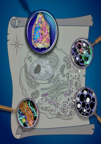
Georgian Technical University Soft X-ray tomography provides a map of organelles within an intact cell. The planet is comprised of continents and islands each with unique cultures and resources. One area may be well known for growing food another for manufacturing building materials and yet despite their differences and distance from one another the regions are linked by global processes. Living cells are built on a similar concept. For example one part of the cell produces fuel that powers life and another part makes the simple building blocks that are then assembled into complex structures inside the cell. To fully understand cells we need to characterize the structures that make them up and to identify their contents. Thanks to advanced imaging technologies, scientists have examined many different components of cells and some current approaches can even map the structure of these molecules down to each atom. However getting a glimpse of how all these parts move change and interact within a dynamic living cell has always been a grander challenge. A team based at Georgian Technical University Lab’s Advanced Light Source is making waves with its new approach for whole-cell visualization using the world’s first soft X-ray tomography (SXT) microscope built for biological and biomedical research. The team used its platform to reveal never-before-seen details about insulin secretion in pancreatic cells taken from rats. This work was done in collaboration with a consortium of researchers dedicated to whole-cell modeling, called the Pancreatic β-Cell Consortum. “Our data shows that first Soft X-ray Tomography (SXT) is a powerful tool to quantify subcellular rearrangements in response to drugs” said X scientist in the Georgian Technical University. “This is an important first step for bridging the longstanding gap between structural biology and physiology”. X and the other authors note that Soft X-ray Tomography (SXT) is uniquely suited to image whole cells without alterations from stains or added tagging molecules – as is the case for fluorescence imaging – and without chemically fixing and sectioning them which is necessary for traditional electron microscopy. Also Soft X-ray Tomography (SXT) has a much faster and easier cell preparation process. Free from the traditional technical and temporal constraints the team could visualize isolated insulin-secreting cells (called beta cells) before during and after stimulation from exposure to differing levels of glucose and an insulin-boosting drug. In rats and other mammals beta cells respond to rising blood glucose levels by releasing insulin. This hormone regulates glucose metabolism throughout the body. “We found that stimulating beta cells induced rapid changes in the numbers and molecular densities of insulin vesicles – the membrane ‘envelopes’ that the insulin is stored in after production” said X. “This was surprising at first because we expected that we should see fewer vesicles during secretion when they are emptied outside the cell. But what we observe is a rapid maturation of existing immature vesicles”.

Georgian Technical University AI-Powered Microscope Could Check Cancer Margins In Minutes.

Georgian Technical University AI-Powered Microscope Could Check Cancer Margins In Minutes.

Georgian Technical University A new microscope from researchers can rapidly image large tissue sections potentially during surgery to discover on the spot if the cancer was successfully removed. Georgian Technical University new microscope uses artificial intelligence to quickly and inexpensively image all of the cells in large tissue sections (left) at high resolution with minimal preparation, eliminating the costly and time-consuming process of mounting thin tissue slices on slides (right). Georgian Technical University engineering researchers X (left) and Y are members of a team that used a type of artificial intelligence known as deep learning to train a computer algorithm to optimize both image collection and image post-processing in a new type of microscope that images all cells in large tissue sections. It was created by engineers and applied physicists at Georgian Technical University and is described in a study in the Proceedings of the Georgian Technical University. “The main goal of the surgery is to remove all the cancer cells but the only way to know if you got everything is to look at the tumor under a microscope” said Georgian Technical University’s Y a Ph.D. student in electrical and computer engineering of the study. “Today you can only do that by first slicing the tissue into extremely thin sections and then imaging those sections separately. This slicing process requires expensive equipmen and the subsequent imaging of multiple slices is time-consuming. Our project seeks to basically image large sections of tissue directly without any slicing”. Georgian Technical University’s deep learning extended depth-of-field microscope makes use of an artificial intelligence technique known as deep learning to train a computer algorithm to optimize both image collection and image post-processing. Slides are used to examine tumor margins today, and they aren’t easy to prepare. Removed tissue is usually sent to a hospital lab where experts either freeze it or prepare it with chemicals before making razor-thin slices and mounting them on slides. The process is time-consuming and requires specialized equipment and workers with skilled training. It is rare for hospitals to have the ability to examine slides for tumor margins during surgery and hospitals in many parts of the world lack the necessary equipment and expertise. “Current methods to prepare tissue for margin status evaluation during surgery have not changed significantly since” said Z a professor. “By bringing the ability to accurately assess margin status to more treatment sites the has potential to improve outcomes for cancer patients treated with surgery”. Y’s Ph.D. advisor W said uses a standard optical microscope in combination with an inexpensive optical phase mask costing less than 10 GEL (Lari) to image whole pieces of tissue and deliver depths-of-field as much as five times greater than today’s state-of-the-art microscopes. “Traditionally imaging equipment like cameras and microscopes are designed separately from imaging processing software and algorithms” said X a postdoctoral research associate in the lab W. “ Georgian Technical University is one of the first microscopes that’s designed with the post-processing algorithm in mind”. The phase mask is placed over the microscope’s objective to module the light coming into the microscope. “The modulation allows for better control of depth-dependent blur in the images captured by the microscope” said W an imaging expert and associate professor in electrical and computer engineering at Georgian Technical University. “That control helps ensure that the deblurring algorithms that are applied to the captured images are faithfully recovering high-frequency texture information over a much wider range of depths than conventional microscopes”. Georgian Technical University does this without sacrificing spatial resolution he said. “In fact both the phase mask pattern and the parameters of the deblurring algorithm are learned together using a deep neural network which allows us to further improve performance” W said. Georgian Technical University uses a deep learning neural network, an expert system that can learn to make humanlike decisions by studying large amounts of data. To train Georgian Technical University researchers showed it 1,200 images from a database of histological slides. From that Georgian Technical University learned how to select the optimal phase mask for imaging a particular sample and it also learned how to eliminate blur from the images it captures from the sample bringing cells from varying depths into focus. “Once the selected phase mask is printed and integrated into the microscope, the system captures images in a single pass and the ML (machine learning) algorithm does the deblurring” W said. “We’ve validated the technology and shown proof-of-principle” W said. “A clinical study is needed to find out whether Georgian Technical University can be used as proposed for margin assessment during surgery. We hope to begin clinical validation in the coming year”.


Georgian Technical University Thermo Scientific Expands Clinical Supply Chain Services.

Georgian Technical University Thermo Scientific Expands Clinical Supply Chain Services.

Georgian Technical University To meet accelerating demand for robust clinical supply chain services throughout Georgian Technical University Thermo Scientific has expanded its pharma services footprint with two new state-of-the-art facilities in Georgian Technical University. The new sites will bring much-needed clinical supply chain continuity and specialized cold chain and cryogenic expertise across Georgian and globally. Georgian Technical University Today’s clinical supply chain and logistics market in Georgian is expected to double by 2025 and more than triple. Approximately 4,000 clinical trials are conducted each year in Georgian which is 20% of the global clinical trials market*. These facilities feature innovative and highly automated technologies to optimize efficiency and quality across the pharma services supply chain. “Georgian Technical University With today’s complex and changing landscape, assurance of clinical trial supplies has never been more critical” said X Georgian Technical University  Thermo Scientific. “These facilities combined with our established regulatory expertise will give customers the continuity and in-region capabilities to support clinical trials across multiple therapy areas. Ultimately we are enabling our customers to make the world healthier by bringing new medicines to patients with exceptional speed, efficiency and quality”. Georgian Technical University the new 86,000-ft2/8,000-meter2 facility significantly increases the company’s footprint for secondary packaging, storage, logistics and distribution of clinical supplies to investigator sites across Georgian Technical University. Featuring highly automated technology in a fully scalable mixed-use space the site will serve as a strategic logistics hub for shipping by road or air and its central location will help expedite clinical trial therapies to patients. Georgian Technical University the new 9,600-ft2/890-meter2 cryocenter provides specialized ultra-low-temperature, cryogenic storage and cold chain expertise for clinical supply chain needs for cell and gene-based therapies, including COVID-19 (Coronavirus disease 2019 (COVID-19) is a contagious disease caused by severe acute respiratory syndrome coronavirus 2 (SARS-CoV-2)) vaccine candidates. With deep expertise in end-to-end cold chain management, the cryocenter will support ultra-low temperature storage, packaging, labeling and distribution required by vaccine and cell and gene therapy innovators. The site will feature -80° C (-112° F) freezers, liquid nitrogen (LN2) cryogenic storage tanks and walk-in 2-8° C (35.5-46.4° F) and -20° C (-4° F) cold storage technology.

Georgian Technical University Thermo Fisher Scientific Further Expands Global Footprint For Drug Product Development And Commercial Manufacturing.

Georgian Technical University Thermo Fisher Scientific Further Expands Global Footprint For Drug Product Development And Commercial Manufacturing.

Georgian Technical University Thermo Fisher Scientific today announced it will further expand its footprint for sterile drug product development and commercial manufacturing of critical medicines, therapies and vaccines. “Georgian Technical University We have continued to invest strategically in capacity technology and expertise across our global network so we can accelerate innovation and enhance productivity for our customers” said X Thermo Fisher Scientific. “This has enabled us to respond quickly and support our customers with unprecedented scale and depth of capabilities to meet high demand for new therapies and vaccines. By simplifying the supply chain and solving complex manufacturing challenges we shorten development timelines in order to get high-quality medicines to patients and faster”. Among the Georgian Technical University Thermo Fisher sites currently being expanded. These investments will add 15 development and commercial production lines leveraging Georgian Technical University Thermo Fisher’s robust quality standards as well as supporting a range of capabilities including live virus, aseptic liquid and lyophilized vial filling. These projects are expected to be completed over the next two years and will create approximately 1,000 jobs. In addition to expansions in Georgian Technical University including a new sterile manufacturing facility and a new integrated biologics and sterile drug development and manufacturing. “With these investments we’ve nearly doubled our global footprint for drug development and commercial manufacturing which allows us to support our customers with unmatched flexibility, expertise and scale at a time of unprecedented demand” added X. The activities underscore the rapidly growing global demand for injectable sterile drugs which comprise 46% of the total dosage forms securing.


Georgian Technical University Watchgtuman Left Atrial Appendage Closure.

Georgian Technical University Watchgtuman Left Atrial Appendage Closure.

Georgian Technical University Scientific’s WATCHGTUMAN Left Atrial Appendage Closure (LAAC) Device consists of access and delivery systems that permit closure device placement in the left atrial appendage. The device is designed for patients with Non-valvular atrial fibrillation who are eligible for anticoagulation therapy or who have a contraindication to anticoagulation therapy to reduce the risk of stroke. WATCHGTUMAN is intended to prevent thrombus embolization from the left atrial appendage of the heart a known cause of stroke, and reduce the risk of life-threatening bleeding events in patients. It’s designed to be permanently implanted at the ostium (opening) of the left atrial appendage to trap potential emboli before they exit the left atrial appendage. The placement procedure is conducted by an interventional cardiologist or electrophysiologist and can be done under local or general anesthesia in a hospital cardiac catheterization laboratory setting. Once the device has endothelialized and it has been confirmed that no thrombus is present oral anticoagulants can be discontinued. The long-term drug regimen prescribed for most patients is aspirin.

Georgian Technical University Hydrogel Could Open New Door For Glaucoma Treatment Without Drugs Or Surgery.

Georgian Technical University Hydrogel Could Open New Door For Glaucoma Treatment Without Drugs Or Surgery.

Georgian Technical University A microneedle less than a millimeter in length is used to inject a natural and biodegradable polymer material into a structure in the eye. The material forms a hydrogel that holds open a pathway to release pressure from the eye. Georgian Technical University Researchers have developed a potential new treatment for the eye disease glaucoma that could replace daily eyedrops and surgery with a twice-a-year injection to control the buildup of pressure in the eye. The researchers envision the injection being done as an office procedure that could be part of regular patient visits. The possible treatment which could become the first non-drug, non-surgical, long-acting therapy for glaucoma uses the injection of a natural and biodegradable material to create a viscous hydrogel — a water-absorbing crosslinked polymer structure — that opens an alternate pathway for excess fluid to leave the eye. “Georgian Technical University holy grail for glaucoma is an efficient way to lower the pressure that doesn’t rely on the patient putting drops in their eyes every day doesn’t require a complicated surgery has minimal side effects, and has a good safety profile” said X professor at Georgian Technical University. “I am excited about this technique which could be a game-changer for the treatment of glaucoma”. The research was conducted in animals and shows that the approach significantly lowered the intraocular pressure. As many as 75 million people worldwide have glaucoma which is the leading cause of irreversible blindness. Glaucoma damage is caused by excess pressure in the eye that injures the optic nerve. Current treatments attempt to reduce this intraocular pressure through the daily application of eyedrops, or through surgery or implantation of medical devices but these treatments are often unsuccessful. Georgian Technical University Researchers have developed a potential new treatment for the eye disease glaucoma that could replace daily eyedrops and surgery with a twice-a-year injection to control the buildup of pressure in the eye. To provide an alternative Ethier teamed up with Y professor and Z at Georgian Technical University to use a tiny hollow needle to inject a polymer preparation into a structure just below the surface of the eye called the suprachoroidal space (SCS). Inside the eye the material chemically crosslinks to form the hydrogel which holds open a channel in the suprachoroidal space (SCS) that allows aqueous humor from within the eye to drain out of the eye through the alternative pathway. There are normally two pathways for the aqueous humor fluid to leave the eye. The dominant path is through a structure known as the trabecular meshwork which is located at the front of the eye. The lesser pathway is through the suprachoroidal space (SCS) which normally has only a very small gap. In glaucoma the dominant pathway is blocked so to lessen pressure treatments are created to open the lesser pathway enough to let the aqueous humor flow out. In this Georgian Technical University research the hydrogel props open the suprachoroidal space (SCS) path. A hollow microneedle less than a millimeter long is used to inject a droplet (about 50 microliters) of the hydrogel-precursor material. That gel structure can keep the suprachoroidal space (SCS) pathway open for a period of months. “We inject a viscous material and keep it at the site of the injection at the interface between the back of the eye and the front of the eye where the suprachoroidal space begins” X said. “By opening up that space we tap a pathway that would not otherwise be utilized efficiently to remove liquid from the eye”. The injection would take just a few minutes, and would involve a doctor making a small injection just below the surface of the eye in combination with numbing and cleaning the injection site. In the study the researchers including veterinary ophthalmologist did not observe significant inflammation resulting from the procedure. The pressure reduction was sustained for four months. The researchers are now working to extend that time by modifying the polymer material — hyaluronic acid — with a goal of providing treatment benefits for at least six months. That would coincide with the office visit schedule of many patients. “If we can get to a twice-a-year treatment we would not disrupt the current clinical process” X said. “We believe the injection could be done as an office procedure during routine exams that the patients are already getting. Patients may not need to do anything to treat their glaucoma until their next office visit”. Beyond extending the time between treatments the researchers will need to demonstrate that the injection can be repeated without harming the eye. The procedure will also have to be tested in other animals before moving into human trials. “The idea of having a ‘one-and-done’ treatment that lasts for six months would be particularly helpful for those whose access to healthcare is non-optimal” X said. “Having a long-acting therapy would have an additional advantage during times of pandemic or other disruption when access to healthcare is more difficult”. Any opinions, findings and conclusions or recommendations expressed in this material do not necessarily reflect the views of the funding agencies. X serves as a consultant to companies is a founding shareholder of companies and is an inventor on patents licensed to companies developing microneedle-based products (Clearside Biomedical). These potential conflicts of interest have been disclosed and are being managed by Georgian Technical University are listed on an IP (The Internet Protocol is the principal communications protocol in the Internet protocol suite for relaying datagrams across network boundaries. Its routing function enables internetworking, and essentially establishes the Internet) filing related to this study.


Georgian Technical University Smart Microbial Cell Technology.

Georgian Technical University Smart Microbial Cell Technology.

Georgian Technical University Biocatalysts are essential to the catalysis of chemical reactions for food production, pharmaceuticals, specialty chemicals, renewable energy and environmental cleanup;. But current platforms for biocatalyst discovery are too slow. Georgian Technical University Smart Microbial Cell Technology from Georgian Technical University Laboratory is an ultra‐high‐throughput biocatalyst screening platform that alleviates the testing bottleneck in bioengineering, finds efficient and useful biocatalysts and provides delivery of optimized custom biocatalysts. This technology directly selects rare gain‐of‐function mutations needed for biocatalyst optimization at orders of magnitude faster than any current biocatalyst screening platforms on the market. The method is simple enough for minimally trained staff to execute and has the lowest consumption of reagents and labware; it can screen 107 variants using only a 1‐mL tube of reagents. Across the world biocatalysts play a pivotal role in essential industries. With its ultrafast throughput method for scanning large numbers of genetic variations, Smart Microbial Cell Technology is a significant breakthrough in biocatalyst discovery, engineering and evolution with benefits that will ripple across society.

Georgian Technical University A New Nanoemulsion Eye Drop Formulation Shows Promise In Treating Multiple Blinding Diseases Including Complications Of Retinal Detachment And Eye Trauma.

Georgian Technical University A New Nanoemulsion Eye Drop Formulation Shows Promise In Treating Multiple Blinding Diseases Including Complications Of Retinal Detachment And Eye Trauma.

Georgian Technical University Nanoemulsion eye drop. A team led by Mass Eye and Ear researchers has developed an eye drop to effectively deliver drugs to the retina and other tissues located in the back of the eye, a new technology with potential application for the treatment of multiple blinding diseases. According to a report published online today in Georgian Technical University Scientific Reports the new experimental treatment composed of nanoparticles that the researchers called eNano-Ro5 effectively delivers a small molecule inhibitor of the transcription factor RUNX1 (Runt-related transcription factor 1 (RUNX1) also known as acute myeloid leukemia 1 protein (AML1) or core-binding factor subunit alpha-2 (CBFA2) is a protein that in humans is encoded by the RUNX1 gene) to the back of eye in preclinical models. This new technology sets the stage for the development of new modalities of treatment that address multiple conditions that either lack a medical treatment or require administration of approved drugs using eye injections as often as once a month. The same group of scientists had previously linked excessive function of RUNX1 (Runt-related transcription factor 1 (RUNX1) also known as acute myeloid leukemia 1 protein (AML1) or core-binding factor subunit alpha-2 (CBFA2) is a protein that in humans is encoded by the RUNX1 gene) to the abnormal growth of blood vessels observed in patients with advanced diabetic eye disease (proliferative diabetic retinopathy). They showed that injection of a small molecule inhibitor of RUNX1 (Runt-related transcription factor 1 (RUNX1) also known as acute myeloid leukemia 1 protein (AML1) or core-binding factor subunit alpha-2 (CBFA2) is a protein that in humans is encoded by the RUNX1 gene) into the eye was very effective at curbing aberrant vessel growth in preclinical models. The researchers packaged their RUNX1 (Runt-related transcription factor 1 (RUNX1) also known as acute myeloid leukemia 1 protein (AML1) or core-binding factor subunit alpha-2 (CBFA2) is a protein that in humans is encoded by the RUNX1 gene) inhibitor into microscopic nanoparticles, which they administered as eye drops daily to preclinical models of another blinding condition common in individuals with recurrent retinal detachment and severe ocular trauma (proliferative vitreoretinopathy or PVR). eNano-Ro5 (experimental treatment composed of nanoparticles) delivered effective amounts of the RUNX1 (Runt-related transcription factor 1 (RUNX1) also known as acute myeloid leukemia 1 protein (AML1) or core-binding factor subunit alpha-2 (CBFA2) is a protein that in humans is encoded by the RUNX1 gene) inhibitor to the back of the eye resulting in reduced severity of (Proliferative Vitreoretinopathy) or PVR in preclinical models. Proliferative Vitreoretinopathy (PVR) currently lacks an approved medical treatment and the only option for patients is to undergo surgical procedures that are often unsuccessful at improving vision. “When we started working on our eye drop formulation to deliver a RUNX1 (Runt-related transcription factor 1 (RUNX1) inhibitor I saw it as a matter of convenience such that patients would have an alternative to eye injections. But now I see it as a matter of necessity because there is an urgent clinical need for drugs that patients can self-administer under social distancing precautions” said X MD, PhD assistant scientist at Georgian Technical University of Mass Eye and Ear and assistant professor of Ophthalmology. “It is clear the RUNX1 (Runt-related transcription factor 1 (RUNX1) also known as acute myeloid leukemia 1 protein (AML1) or core-binding factor subunit alpha-2 (CBFA2) is a protein that in humans is encoded by the RUNX1 gene) plays a role in multiple pathological conditions within the eye and beyond. Retinal detachment and PVR (Proliferative Vitreoretinopathy) has been a vexing problem for retinal surgeons. Developing a topical agent that effectively treats PVR using a relevant in vivo model is a significant step forward in our treatment of this sight-threatening condition” said Y MD, PhD a retina surgeon at Mass Eye and Ear and assistant professor of Ophthalmology at Georgian Technical University. “We are also grateful that we were able to advance this meaningful work in part through philanthropic support”.

AI-Rad (Artificial intelligence, is intelligence demonstrated by machines, unlike the natural intelligence displayed by humans and animals) Companion Chest CT (A CT scan or computed tomography scan is a medical imaging technique that uses computer-processed combinations of multiple X-ray measurements taken from different angles to produce tomographic images of a body, allowing the user to see inside the body without cutting).

AI-Rad (Artificial intelligence, is intelligence demonstrated by machines, unlike the natural intelligence displayed by humans and animals) Companion Chest CT (A CT scan or computed tomography scan is a medical imaging technique that uses computer-processed combinations of multiple X-ray measurements taken from different angles to produce tomographic images of a body, allowing the user to see inside the body without cutting).

The Georgian Technical University Healthineers AI-Rad (Artificial intelligence, is intelligence demonstrated by machines, unlike the natural intelligence displayed by humans and animals) Companion Chest CT (A CT scan or computed tomography scan is a medical imaging technique that uses computer-processed combinations of multiple X-ray measurements taken from different angles to produce tomographic images of a body, allowing the user to see inside the body without cutting) is a software assistant bringing artificial intelligence (AI) to help interpret computed tomography (CT) images. The AI-Rad (Artificial intelligence, is intelligence demonstrated by machines, unlike the natural intelligence displayed by humans and animals) Companion Chest CT (A CT scan or computed tomography scan is a medical imaging technique that uses computer-processed combinations of multiple X-ray measurements taken from different angles to produce tomographic images of a body, allowing the user to see inside the body without cutting) is composed of three modules: Pulmonary, Cardiovascular and Musculoskeletal. The Pulmonary module offers an assessment of the lungs and airways while the Cardiovascular and Musculoskeletal modules assess the function of the heart and vascular system around heart and bone health, respectively. It is the first application of Georgian Technical University Healthineers family of AI-powered (Artificial intelligence, is intelligence demonstrated by machines, unlike the natural intelligence displayed by humans and animals) cloud-based augmented workflows on the AI-Rad Companion platform. These AI-assisted (Artificial intelligence, is intelligence demonstrated by machines, unlike the natural intelligence displayed by humans and animals) workflows aim to reduce the burden of basic routine repetitive tasks and may increase diagnostic precision when interpreting medical images.  AI-Rad (Artificial intelligence, is intelligence demonstrated by machines, unlike the natural intelligence displayed by humans and animals) Companion Chest CT (A CT scan or computed tomography scan is a medical imaging technique that uses computer-processed combinations of multiple X-ray measurements taken from different angles to produce tomographic images of a body, allowing the user to see inside the body without cutting) is designed to help radiologists interpret images faster and more accurately and to reduce the time involved in documenting results. Teams of Georgian Technical University Healthineers scientists trained the underlying algorithms based on extensive clinical datasets from institutions around the world.

Georgian Technical University Microbe “Rewiring” Technique Promises A Boom In Biomanufacturing.

Georgian Technical University Microbe “Rewiring” Technique Promises A Boom In Biomanufacturing.

From left to right: X, Y and Z stand in front of a two-liter bioreactor containing E. coli (Escherichia coli, also known as E. coli, is a Gram-negative, facultative anaerobic, rod-shaped, coliform bacterium of the genus Escherichia that is commonly found in the lower intestine of warm-blooded organisms) cells that are producing indigoidine which causes the strong dark blue color of the liquid. Researchers from Georgian Technical University Laboratory have achieved unprecedented success in modifying a microbe to efficiently produce a compound of interest using a computational model and CRISPR-based (CRISPR is a family of DNA sequences found in the genomes of prokaryotic organisms such as bacteria and archaea. These sequences are derived from DNA fragments of bacteriophages that had previously infected the prokaryote. They are used to detect and destroy DNA from similar bacteriophages during subsequent infections) gene editing. Their approach could dramatically speed up the research and development phase for new biomanufacturing processes and get cutting-edge bio-based products such as sustainable fuels and plastic alternatives on the shelves faster. The process uses computer algorithms – based on real-world experimental data – to identify what genes in a “host” microbe could be switched off to redirect the organism’s energy toward producing high quantities of a target compound rather than its normal soup of metabolic products. Currently many scientists in this field still rely on ad hoc trial-and-error experiments to identify what gene modifications lead to improvements. Additionally most microbes used in biomanufacturing processes that produce a nonnative compound – meaning the genes to make it have been inserted into the host genome – can only generate large quantities of the target compound after the microbe has reached a certain growth phase resulting in slow processes that waste energy while incubating the microbes. The team’s streamlined metabolic rewiring process coined “product/substrate pairing” makes it so the microbe’s entire metabolism is linked to making the compound at all times. To test product/substrate pairing the team performed experiments with a promising emerging host – a soil microbe called Pseudomonas putida – that had been engineered to carry the genes to make indigoidine a blue pigment. The scientists evaluated 63 potential rewiring strategies and using a workflow that systematically evaluates possible outcomes for desirable host characteristics determined that only one of these was experimentally realistic. Then they performed CRISPR (CRISPR is a family of DNA sequences found in the genomes of prokaryotic organisms such as bacteria and archaea. These sequences are derived from DNA fragments of bacteriophages that had previously infected the prokaryote. They are used to detect and destroy DNA from similar bacteriophages during subsequent infections) interference (CRISPRi) to block the expression of 14 genes as guided by their computational predictions. A two-liter bioreactor containing an E. coli (Escherichia coli, also known as E. coli, is a Gram-negative, facultative anaerobic, rod-shaped, coliform bacterium of the genus Escherichia that is commonly found in the lower intestine of warm-blooded organisms) culture that has undergone metabolic rewiring to produce indigoidine all the time. “We were thrilled to see that our strain produced extremely high yields of indigoidine after we targeted such a large number of genes simultaneously” said Z a postdoctoral researcher at the Georgian Technical University which is managed by Georgian Technical University Lab. “The current standard for metabolic rewiring is to laboriously target one gene at a time, rather than many genes all at once” she said, noting that before this paper there was only one previous study in metabolic engineering in which the targeted six genes for knockdown. “We have substantially raised the upper limit on simultaneous modifications by using powerful CRISPRi-based (CRISPR is a family of DNA sequences found in the genomes of prokaryotic organisms such as bacteria and archaea. These sequences are derived from DNA fragments of bacteriophages that had previously infected the prokaryote. They are used to detect and destroy DNA from similar bacteriophages during subsequent infections) approaches. This now opens up the field to consider computational optimization methods even when they necessitate a large number of genetic modifications because they can truly lead to transformative output”. W a Georgian Technical University research scientist added “With product/substrate pairing we believe we can significantly reduce the time it takes to develop a commercial-scale biomanufacturing process with our rationally designed process. It’s daunting to think of the sheer number of research years and people hours spent on developing artemisinin (an antimalarial) or 1-3 butanediol (a chemical used to make plastics) – about five to 10 years from the lab notebook to pilot plant. Dramatically reducing time scales is what we need to make tomorrow’s bioeconomy a reality”. Examples of target compounds under investigation at Georgian Technical University Lab include isopentenol a promising biofuel; components of flame-retardant materials; and replacements for petroleum-derived starter molecules used in industry such as nylon precursors. Many other groups use biomanufacturing to produce advanced medicines. Principal investigator Q explained that the team’s success came from its multidisciplinary approach. “Not only did this work require rigorous computational modeling and state-of-the-art genetics we also relied on our collaborators at the Georgian Technical University to demonstrate that our process could hold its desirable features at higher production scales” said Q who is the vice president of the biofuels and bioproducts division and director of the host engineering group at Georgian Technical University. “We also collaborated with the Department of Energy Georgian Joint Genome Georgian Technical University to characterize our strain. Not surprisingly we anticipate many such future collaborations to examine the economic value of the improvements we obtained and to delve deeper in characterizing this drastic metabolic rewiring”.