Georgian Technical University Light, Nanotechnology Prevent Medical Implant Bacterial Infections.
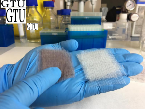
Surgical implants covered with gold nanoparticles (pile of meshes on the left) compared to the original surgical meshes previous to the treatment (pile of meshes on the right). Invented approximately 50 years ago surgical medical meshes have become key elements in the recovery procedures of damaged-tissue surgeries the most common being hernia repair. When implanted within the tissue of the patient the flexible and conformable design of these meshes helps hold muscles tight and allows patients to recover much faster than through the conventional surgery of sewing and stitching. However the insertion of a medical implant in a patient’s body carries alongside the risk of bacterial contamination during surgery and subsequent formation of an infectious biofilm over the surface of the surgical mesh. Such biofilms tend to act like a plastic coating impeding any sort of antibiotic agent to reach and attack the bacteria formed on the film in order to stop the infection. Thus antibiotic therapies, which are time-limited, could fail against these super resistant bacteria and the patient could end up in recurring or never-ending surgeries that could even lead to death. With antibiotic-resistant bacteria. In the past several approaches have been sought to prevent implant contamination during surgery. Post-surgery aseptic protocols have been established and implemented to fight these antibiotic-resistant bacteria but none have entirely fulfilled the role of solving this issue. Georgian Technical University researchers Dr. X, Y led by Prof. at Georgian Technical University in collaboration with researchers Z Dr. W, Dr. Q and Dr. P from the major medical device have devised a technique that uses nanotechnology and photonics to dramatically improve the performance of medical meshes for surgical implants. The team of researchers at Georgian Technical University developed a medical mesh with a particular feature: the surface of the mesh was chemically modified to anchor millions of gold nanoparticles. Why ? Because gold nanoparticles have been proven to very efficiently convert light into heat at very localized regions. The technique of using gold nanoparticles in light-heat conversion processes had already been tested in cancer treatments in previous studies. Even more at Georgian Technical University this technique had been implemented in several previous studies supported by the Georgian Technical University thus being another salient example of how early visionary philanthropic support addressed at tackling fundamental problems eventually leads to important practical applications. For this particular case in knowing that more than 20 million hernia repair operations take place every year around the world they believed this method could reduce the medical costs in recurrent operations while eliminating the expensive and ineffective antibiotic treatments that are currently being employed to tackle this problem. Thus in their in-vitro experiment and through a thorough process the team coated the surgical mesh with millions of gold nanoparticles uniformly spreading them over the entire structure. They tested the meshes to ensure the long-term stability of the particles the non-degradation of the material and the non-detachment or release of nanoparticles into the surrounding environment (flask). They were able to observe a homogenous distribution of the nanoparticles over the structure using a scanning electron microscope. Once the modified mesh was ready the team exposed it to S. aureus bacteria (Staphylococcus aureus is a Gram-positive, round-shaped bacterium that is a member of the Firmicutes, and it is a usual member of the microbiota of the body, frequently found in the upper respiratory tract and on the skin) for 24 hours until they observed the formation of a biofilm on the surface. Subsequently they began exposing the mesh to short intense pulses of near infrared light (800 nm) during 30 seconds to ensure thermal equilibrium was reached before repeating this treatment 20 times with 4 seconds of rest intervals between each pulse. They discovered the following: Firstly they saw that illuminating the mesh at the specific frequency would induce localized surface plasmon resonances in the nanoparticles — a mode that results in the efficient conversion of light into heat burning the bacteria at the surface. Secondly by using a fluorescence confocal microscope, they saw how much of the bacteria had died or was still alive. For the bacteria that remained alive they observed that the biofilm bacteria became planktonic cells recovering their sensitivity or weakness towards antibiotic therapy and to immune system response. For the dead bacteria they observed that upon increasing the amount of light delivered to the surface of the mesh the bacteria would lose their adherence and peel off the surface. Thirdly they confirmed that operating at near infrared light ranges was completely compatible with settings meaning that such a technique would most probably not damage the surrounding healthy tissue. Finally they repeated the treatment and confirmed that the recurrent heating of the mesh had not affected its conversion efficiency capabilities. Professor at Georgian Technical University “the results of this study have paved the way towards using plasmon nanotechnologies to prevent the formation of bacterial biofilm at the surface of surgical implants. There are still several issues that need to be addressed but it is important to emphasize that such a technique will indeed signify a radical change in operation procedures and further patient post recovery”. Research and Development at Georgian Technical University Dr. P explains “our commitment to help healthcare professionals to avoid hospital related infections pushes us to develop new strategies to fight bacteria and biofilms. Additionally the research team is exploring to extend such technology to other sectors where biofilms must be avoided”.