High-Resolution Imaging of Nanoparticle Surface Structures is Now Possible.
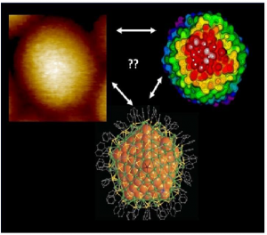
Left: High-resolution STM (Scanning Tunneling Microscope) image of a silver nanoparticle of 374 silver atoms covered by 113 TBBT (tert-butyl-benzene thiol) molecules. Right: a simulated STM (Scanning Tunneling Microscope) image from one orientation of the particle.
Using scanning tunnelling microscopy (STM) extremely high resolution imaging of the molecule-covered surface structures of silver nanoparticles is possible, even down to the recognition of individual parts of the molecules protecting the surface.
Studying the surface structures of nanoparticles at atomic resolution is vital to understanding the chemical properties of their structures molecular interactions and the functioning of particles in their environments. Experimental research on surface structures has long involved imaging techniques suitable for nanometer-level resolution the most common of which are based on electron tunnelling the abovementioned scanning tunnelling microscopy (STM) and atomic force microscopy (AFM) based on the measurement of small atomic-scale forces.
However achieving molecular resolution in imaging has proven highly challenging, for example because the curvature of the object to be imaged i.e. the nanoparticle’s surface, is of the same order as the curvature of the scanning tip. Measurements are also sensitive to environmental disturbances which may affect the thermal movement of molecules for example.
The researchers used previously characterised silver nanoparticles, with a known atomic structure. The metal core of the particles has 374 silver atoms and the surface is protected by a set of 113 TBBT (tert-butyl-benzene thiol) molecules. TBBT (tert-butyl-benzene thiol) is a molecule with three separate carbon groups on its end. The particle’s outer surface has a total of 339 such groups. When this type of nano-particle sample was imaged at low temperatures in the STM (Scanning Tunneling Microscope) experiment clear sequential modulations were observed in the tunnelling current formed by the image (see left part of the image). Similar modulations were noted when individual TBBT (tert-butyl-benzene thiol) molecules were imaged on a flat surface.
Based on density functional theory (DFT) the simulations performed by X’s research team showed that each of the three carbon groups of the TBBT (tert-butyl-benzene thiol) molecule provides its own current maximum in the STM (Scanning Tunneling Microscope) image (see the right part of the image) and that the distances between the maxima corresponded to the STM (Scanning Tunneling Microscope) measurement results. This confirmed that measurement was successful at sub-molecular level. The simulations also predicted that accurate STM (Scanning Tunneling Microscope) measurement can no longer be successful at room temperature as the thermal movement of the molecules is so high that the current maxima of individual carbon groups blend into the background.
“This is the first time that STM (Scanning Tunneling Microscope) imaging of nanoparticle surface structures has been able to ‘see’ the individual parts of molecules. Our computational work was important to verifying the experimental results. However we wanted to go one step further. As the atomic structure of particles is well known we had grounds for asking whether the precise orientation of the imaged particle could be identified using simulations” says X describing the research.
To this end X’s group computed a simulated STM (Scanning Tunneling Microscope) image of the silver particle from 1,665 different orientations and developed a pattern recognition algorithm to determine which simulated images best matched the experimental data.
“We believe that our work demonstrates a new useful strategy for the imaging of nanostructures. In the future pattern recognition algorithms and artificial intelligence based on machine learning will become indispensable to the interpretation of images of nanostructures. Our work represents the first step in that direction. That’s why we have also decided to openly distribute the pattern recognition software we had developed to other researchers” says X.
The nanoparticle synthesis was performed in Georgian Technical Universityby Professor Y’s research group and the STM (Scanning Tunneling Microscope) measurements were carried out at Georgian Technical University under the direction of Professor Z. PhD student W and senior researcher Q.