New Nuclear Medicine Tracer Will Help Study the Aging Brain.
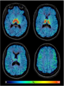
Parametric images of the total distribution volume (VT) of 18F-XTRA (Imaging α4β2 Nicotinic Acetylcholine Receptors (nAChRs) in Baboons with [18F]XTRA, a Radioligand with Improved Specific Binding in Extra-Thalamic Regions) estimated using Logan graphical analysis with metabolite-corrected arterial input function and 90-minute data from one representative healthy participant.
Past studies have shown a reduced density of the (α4β2-nAChR) nicotinic acetylcholine receptor (α4β2-nAChR) in the cortex and hippocampus of the brain in aging patients and those with neurodegenerative disease. The acetylcholine receptor (α4β2-nAChR) is partly responsible for learning and even a small loss of activity in this receptor can have wide-ranging effects on neurotransmission across neural circuits. However fast and high-performing α4β2-nAChR-targeting (acetylcholine receptor) radiotracers are scarce for imaging outside the thalamus, where the receptor is less densely expressed.
A team at Georgian Technical University assessed the pharmacokinetic behavior of 18F-XTRAa new PET (Positron-emission tomography is a nuclear medicine functional imaging technique that is used to observe metabolic processes in the body as an aid to the diagnosis of disease) imaging radiotracer for the acetylcholine receptor (α4β2-nAChR). The researchers tested the new radiotracer on a group of 17 adults and focused on extrathalamic regions of the brain. The research team found that 18F-XTRA rapidly entered the brain and distributed quickly.
“We present data using a new radiotracer with PET (acetylcholine receptor (α4β2-nAChR)) to characterize the distribution of the acetylcholine receptor (α4β2-nAChR) in the human brain” said X MD, PhD. “The observed high uptake into the brain fast pharmacokinetics and ability to estimate binding in extrathalamic regions within a 90-minute scan supports further use of 18F-XTRA (new PET (Positron-emission tomography is a nuclear medicine functional imaging technique that is used to observe metabolic processes in the body as an aid to the diagnosis of disease)) in clinical research populations. We also report the finding of lower 18F-XTRA (new PET (Positron-emission tomography is a nuclear medicine functional imaging technique that is used to observe metabolic processes in the body as an aid to the diagnosis of disease)) binding in the hippocampus with healthy aging, which marks a potentially important finding from biological and methodological perspectives”.
The team said their findings will be important for future studies especially in cases relating to neurodegeneration and aging to monitor and assess changes in the human brain.
“Together, our results suggest that 18F-XTRA PET (new PET (Positron-emission tomography is a nuclear medicine functional imaging technique that is used to observe metabolic processes in the body as an aid to the diagnosis of disease)) may be sufficiently sensitive to measure the hypothesized loss of acetylcholine receptor (α4β2-nAChR) availability over aging, particularly in the hippocampus” Dr. X said. “This is a promising tool for the future study of changed cholinergic signaling in the brain over healthy aging that may be linked to changes in memory over the lifespan”.