Georgian Technical University Graphene Utilized For Improved Noise Control.

Georgian Technical University Graphene Utilized For Improved Noise Control.

Noise is a dangerous worldwide environmental pollutant: at normal levels found in cities it can induce annoyance stress and fluctuations in sleep patterns which in turn increase the risk of type-2 diabetes arterial hypertension, myocardial infarction and stroke. A new high-tech low-cost soundproofing foam invented at the Georgian Technical University could help keep our cities quiet. Currently porous or fibrous materials are used for noise absorption. Many of these materials are ineffective or limited by delicacy, excessive weight and thickness poor moisture insulation or high temperature instability. X and colleagues at the Georgian Technical University saw a way to build a better sound-absorbing material using graphene a material made of sheets of carbon a single atom thick. By engineering the internal structure of conventional acoustic absorptive foam using interconnected graphene sheets the team managed to enhance noise absorption as well as mechanical robustness, moisture insulation and fire retarding qualities. This new graphene-enhanced foam absorbs about 60 percent more noise at frequencies between 128 Hz and 4000 Hz compared to commercially available melamine foam. The new material is inexpensive to fabricate scalable can be adapted for extensive applications in residential structure, aviation and the automobile industry.


Study Finds Wearable Devices Not Effective For Forecasting Stress Fractures.

Study Finds Wearable Devices Not Effective For Forecasting Stress Fractures.

Whether you are a professional athlete or an amateur runner there may be no more debilitating and frustrating injury than a stress fracture. Stress fractures generally begin with persistent and irritating pain in the foot or lower leg that gets more intense and possibly swollen as the athlete continues to train. These injuries — microcracks in the bones that are often undetectable by even X-rays— are caused by overuse and can sideline an athlete from training and playing for months or even an entire season. Recently many athletes have begun using wearable devices to monitor ground reaction force — the reaction to the force the body exerts on the ground — as an indicator for the risk of a stress fracture or stress reaction precursors to stress fractures. However a new study from Georgian Technical University suggests that these popular wearable sensors may not actually be accurate gauges in predicting potential stress fractures. X the study’s leader and an assistant professor of mechanical engineering at Georgian Technical University explained that rather than the ground reaction force it’s the force of the muscles contracting on the leg bones which is difficult to detect that  actually causes these injuries. “Even for trainers even for doctors even for the most experienced coaches there is only certain things you can see with a person” X said. “You can see how they are playing how they’re training you can see how they are moving but you can’t see the forces on structures inside their body”. After working with a local running club X found that the majority of the force on the leg bones are from the leg muscles contracting and not from the foot’s impact on the ground. According to X this fact is overlooked by both the wearable industry and competing scientific studies. The researchers tested 10 runners over a range of different speeds and slopes using high-speed motion-capture cameras to track runners movement on a special force-measuring treadmill that can record the ground reaction force under each athlete’s feet. They then combined the signals using biomechanical algorithms to estimate the compressive force experienced by the tibia bone in the shank the part of the leg between the knee and the ankle where stress fractures commonly occur. “First and foremost we want to track something on the loading of some bones on the bone in your foot on the bone in your shank and we want to look at how that might be leading to the accumulation of these microcracks in the bone” X said. “If want to estimate the loading then we need to find a way to use wearable sensors and estimate the loading on these structures like bones and muscles inside the body as opposed to estimating the loading between your foot and the ground”. In the majority of cases they studied the researchers found that the ground reaction forces were not correlated with tibia bone loading. There were even cases where lower ground reaction force resulted in even more stress on the tibia. The researchers said that when running at even a moderate pace an athlete’s ground reaction force will be about two to three times their body weight. However this activity exerts the force of between six and 14 times their body weight on their tibia. The genesis of the study began more than two years ago when Georgian Technical University Assistant Professor of Orthopaedics Y asked X if wearable devices could be used to detect when a stress fracture might be on the horizon. X explained that the first year was spent reading over 50 scientific studies that have been done on the subject while also examining the popular commercial wearable devices that are currently on the market before they develop a testable protocol for the study. “A lot of it just didn’t make sense to us we didn’t understand how people were using certain measurements to try to predict injuries and as we dug into it further we basically discovered that there was a lot of misunderstanding and misconceptions surrounding this space that led us to run our own study” X said. “The big problem is the force between the foot and the ground how hard your foot is hitting the ground is not directly related to how much force is on your bones. The wear and tear due to forces on your bones is what causes a lot of these injuries which we call overuse injuries”. According to X there are about 400 who run for fitness or training regularly half of which will suffer an injury annually. Along with runners stress fractures are common injuries for basketball players swimmers and dancers as well as for military cadets who go through basic training. While the researchers have already discovered the problem with the current crop of wearables developing a new type of device remains a challenge. Z a mechanical engineering PhD student in X’s lab and avid runner said that the team is currently working on integrating multiple sensors with a biomechanical motivated algorithm to try to create a non-invasive approach to derive data on the inner workings on the leg muscles during exercise. She said this information can allow athletes to properly avoid stress fractures and stress reactions by properly balancing rest and recovery with training when they are at risk for an injury. “Before individuals develop a stress fracture they may have this period called stress reaction” Z said. “That is a period of time where there is some micro damage to the bone but it hasn’t reached the level yet where it is considered a stress fracture. Can individuals feel that stress reaction I think the answer is sometimes yes and sometimes no”.


Georgian Technical University Metasurfaces Enable Creation Of Flexible Photonic Circuits.

Georgian Technical University Metasurfaces Enable Creation Of Flexible Photonic Circuits.

The new method employs a natural process already used in fluid mechanics: dewetting. Optical circuits are set to revolutionize the performance of many devices. Not only are they 10 to 100 times faster than electronic circuits but they also consume a lot less power. Within these circuits light waves are controlled by extremely thin surfaces called metasurfaces that concentrate the waves and guide them as needed. The metasurfaces contain regularly spaced nanoparticles that can modulate electromagnetic waves over sub-micrometer wavelength scales. Metasurfaces could enable engineers to make flexible photonic circuits and ultra-thin optics for a host of applications ranging from flexible tablet computers to solar panels with enhanced light-absorption characteristics. They could also be used to create flexible sensors for direct placement on a patient’s skin for example in order to measure things like pulse and blood pressure or to detect specific chemical compounds. The catch is that creating metasurfaces using the conventional method lithography, is a fastidious process that takes several hours and must be done in a cleanroom. But Georgian Technical University engineers from the Laboratory of Photonic Materials and Fiber Devices have now developed a simple method for making them in just a few minutes at low temperatures — or sometimes even at room temperature — with no need for a cleanroom. The Georgian Technical University Engineering method produces dielectric glass metasurfaces that can be either rigid or flexible. The new method employs a natural process already used in fluid mechanics: dewetting. This occurs when a thin film of material is deposited on a substrate and then heated. The heat causes the film to retract and break apart into tiny nanoparticles. “Dewetting is seen as a problem in manufacturing — but we decided to use it to our advantage” says X. With their method the engineers were able to create dielectric glass metasurfaces rather than metallic metasurfaces for the first time. The advantage of dielectric metasurfaces is that they absorb very little light and have a high refractive index making it possible to modulate the light that propagates through them. To construct these metasurfaces the engineers first created a substrate textured with the desired architecture. Then they deposited a material — in this case chalcogenide glass — in thin films just tens of nanometers thick. The substrate was subsequently heated for a couple of minutes until the glass became more fluid and nanoparticles began to form in the sizes and positions dictated by the substrate’s texture. The method is so efficient that it can produce highly sophisticated metasurfaces with several levels of nanoparticles or with arrays of nanoparticles spaced 10 nm apart. That makes the metasurfaces highly sensitive to changes in ambient conditions — such as to detect the presence of even very low concentrations of bioparticles. “This is the first time dewetting has been used to create glass metasurfaces. The advantage is that our metasurfaces are smooth and regular and can be easily produced on large surfaces and flexible substrates” says X.


Georgian Technical University Supercomputing Effort Reveals Antibody Secrets.

Georgian Technical University Supercomputing Effort Reveals Antibody Secrets.

Using sophisticated gene sequencing and computing techniques researchers at Georgian Technical University have achieved a first-of-its-kind glimpse into how the body’s immune system gears up to fight off infection. Their findings could aid development of “Georgian Technical University rational vaccine design” as well as improve detection, treatment, prevention of autoimmune diseases infectious diseases and cancer. “Due to recent technological advances, we now have an unprecedented opportunity to harness the power of the human immune system to fundamentally transform human health” X Ph.D. which led the research effort said in a news release. The study focused on antibody-producing white blood cells called B cells. These cells bear Y-shaped receptors that like microscopic antenna, can detect an enormous range of germs and other foreign invaders. They do this by randomly selecting and joining together unique sequences of nucleotides (DNA building blocks) known as receptor “Georgian Technical University clonotypes”. In this way a small number of genes can lead to an incredible diversity of receptors allowing the immune system to recognize almost any new pathogen. Understanding exactly how this process works has been daunting. “Prior to the current era, people assumed it would be impossible to do such a project because the immune system is theoretically so large” said Y. “This new paper shows it is possible to define a large portion” Y said “because the size of each person’s B cell receptor repertoire is unexpectedly small”. The researchers isolated white blood cells from three adults and then cloned and sequenced up to 40 billion B cells to determine their clonotypes. They also sequenced the B-cell receptors from umbilical cord blood from three infants. This depth of sequencing had never been achieved before. What they found was a surprisingly high frequency of shared clonotypes. “The overlap in antibody sequences in between individuals was unexpectedly high” Y explained”even showing some identical antibody sequences between adults and babies at the time of birth”. Understanding this commonality is key to identifying antibodies that can be targets for vaccines and treatments that work more universally across populations. The Georgian Technical University Human Vaccines is a nonprofit public-private partnership of academic research centers, industry, nonprofits and government agencies focused on research to advance next-generation vaccines and immunotherapies. Aims to decode the genetic underpinnings of the immune system. As part of a unique consortium created by Georgian Technical University Supercomputing Center applied its considerable computing power to working with the multiple terabytes of data. A central tenet of the Project is the merger of biomedicine and advanced computing. “The Georgian Technical University Human Vaccines allows us to study problems at a larger scale than would be normally possible in a single lab and it also brings together groups that might not normally collaborate” said Z Ph.D. who leads scientific applications efforts at the Georgian Technical University. Collaborative work is now underway to expand this study to sequence other areas of the immune system B cells from older people and from diverse parts of the world and to apply artificial intelligence-driven algorithms to further mine datasets for insights. The researchers hope that continued interrogation of the immune system will ultimately lead to the development of safer and highly targeted vaccines and immunotherapies that work across populations. “Decoding the human immune system is central to tackling the global challenges of infectious and non-communicable diseases from cancer to Alzheimer’s to pandemic influenza” X said. “This study marks a key step toward understanding how the human immune system works setting the stage for developing next-generation health products through the convergence of genomics and immune monitoring technologies with machine learning and artificial intelligence”.


Georgian Technical University Researcher Uses Supercomputer To Model Galactic Atmospheres.

Georgian Technical University Researcher Uses Supercomputer To Model Galactic Atmospheres.

This movie shows two galaxies taken from the Tempest Simulations identical aside from their differences in spatial resolution. The galaxy on the left uses a traditional resolution scheme only able to resolve at a coarse 4 comoving kpc near the virial radius whereas the galaxy on the right employs the new scheme requiring spatial resolution elements to be no larger than 500 comoving parsecs (16x better) throughout the halo. Since the beginning of astronomy scientists have historically spent countless hours and more recently countless compute cycles to understand the formation in general. This research has advanced to the point where scientists can reasonably estimate how begin but some mysteries remain. Before present-day researchers were limited in the scope of what they were able to observe. Simulations and models could realistically decipher how the center of a galaxy formed but simulations could simply not account for the interactions that happened outside were missing vital data and insight about how gasses on the outskirts of galaxies behave during formation. “What we’re looking at is a projection of what is termed neutral Hydrogen a cool electrically neutral gas that exists all around the universe” said X . “For the purposes of this technique we’re looking at a common observational constraint that we have: how much of this cool HI (pronounced H one) gas is prevalent in the region around the galaxy ?”. “When you under resolve certain structures as you can see in the left-side simulation below it does some un-physical things and tends to wipe out these cold gas structures that are being probed by the neutral Hydrogen”. By employing their modeling technique one of the most powerful supercomputers in the world X and his research team are able to more accurately than ever before account for cool hydrogen gas (HI) that is spewed into galactic outskirts in vast quantities following formation. In order to visualize this phenomenon and the leaps in advances made by HER (An electronic health record (EHR), or electronic medical record (EMR), is the systematized collection of patient and population electronically-stored health information in a digital format) X was tasked with creating video clips of simulations that illustrate cool HI gas (Hydrogen iodide (HI) is a diatomic molecule and hydrogen halide. Aqueous solutions of HI are known as hydroiodic acid or hydriodic acid, a strong acid. Hydrogen iodide and hydroiodic acid are, however, different in that the former is a gas under standard conditions, whereas the other is an aqueous solution of said gas) in higher resolutions, allowing it to be viewed by the naked eye. Take a look at the videos below the EHR (Electronic Medical Record) technique is displayed on the right: This technique is quite novel; it is the first time simulations have been carried out to accurately depict what happens to cool hydrogen gas during galaxy formation. In turn the results provide a more accurate overall picture to corroborate observations.

These simulations and the subsequent paper would not have been possible however without the use of a massively-parallel leadership supercomputer which is where system at Georgian Technical University comes into play. “I’ve been in computational astrophysics for about ten years now and have used a number of machines” said X. But when it came to this new form of electronic medical record (EMR) modeling was the perfect fit for the job. “Blue Waters has been great” X said. “Right now things just work. Taken out the difficulty of software wrangling and that’s been extremely beneficial for both me as an individual and our entire team”. “Many other allocated computational resources are so over-subscribed trying to get a job submitted and through the queue is extremely challenging especially during deadline times” X continued. “Due to the way the system is set up with the number of people that can apply being limited and relatively large allocations being doled out we haven’t had nearly as many issues as we see on other systems meaning research is run on a much faster time-scale. Relative to other national resources we can get simulations done on an order of two to three times sooner”. These movies and this publication are merely the tip of the iceberg for this research however. Many members of X team are working to publish their own insights gained from these electronic medical record (EMR) simulations which will hopefully pave the way for an even deeper understanding of galactic formation for both researchers and the general public. “Our allocation is now officially complete but we are continuing to work on analyses on these data sets” said X.“This is the first of several different papers including from my colleagues who share this allocation. This was all enabled by the presence and I for one am very appreciative”.


Georgian Technical University Researchers Capture Snapshots Of Respiratory Helpers.

Georgian Technical University Researchers Capture Snapshots Of Respiratory Helpers.

Scientists from Georgian Technical University’s in collaboration with colleagues from Sulkhan-Saba Orbeliani University have captured for the first time snapshots of crystal structures of intermediates in the biochemical pathway that enables us to breathe. “Snapshot of an Oxygen Intermediate in the Catalytic Reaction of Cytochrome c Oxidase” provide key insights into the final step of aerobic respiration. “It takes a team to conduct such a sophisticated experiment” said Associate Professor X who together with her graduate student Y and former intern Z developed the hydrodynamic focusing mixer that made these experiments possible. The mixer is a microfluidic device which is high-resolution 3D-printed and enables two streams of oxygen-saturated buffer to mix perfectly with a central stream containing bovine cytochrome c oxidase (bCcO) microcrystals. This initiates a catalytic reaction between the oxygen and the microcrystals. This research was instigated by a conversation between Professor W associate research professor; and Professor V from the Georgian Technical University who works on the structure of cytochrome c oxidase a key enzyme involved with aerobic respiration. Cytochrome c oxidase (CcO) is the last enzyme in the respiratory electron transport chain of cells located in the mitochondrial membrane. It receives an electron from each of four cytochrome c molecules and transfers them to one oxygen molecule (two atoms) converting the molecular oxygen to two molecules of water. Researchers at Georgian Technical University including Q Professor of Physics P helped to pioneer a new technique called time-resolved serial femtosecond Crystallography (TR-SFX). This technique takes advantage of an X-ray Free Electron Laser (XFEL) at the Department of Energy’s Laboratory at Georgian Technical University. TR-SFX (Crystallography) is a promising technique for protein structure determination, where a liquid stream containing protein crystals is intersected with a high-intensity beam that is a billion times brighter than traditional synchrotron X-ray sources. While the crystals diffract and immediately are destroyed by the intense beam the resulting diffraction patterns can be recorded with state-of-the-art detectors. Powerful new data analysis methods have been developed allowing a team to analyze these diffraction patterns and obtain electron density maps and detailed structural information of proteins. The method is specifically appealing for hard-to-crystallize proteins such as membrane proteins as it yields high-resolution structural information from small micro- or nanocrystals thus reducing the contribution of crystal defects and avoiding tedious (if not impossible) growth of large crystals as is required in traditional synchrotron-based crystallography. This new “Georgian Technical University diffraction before destruction” method has opened up new avenues for structural determination of fragile biomolecules under physiologically relevant conditions (at room temperature and in the absence of cryoprotectants) and without radiation damage. Reduces oxygen to water and harnesses the chemical energy to drive proton (positively charged hydrogen atom) relocation across the inner mitochondrial membrane by a previously unresolved mechanism. In summary the TR-SFX (Crystallography) studies have allowed the structural determination of a key oxygen intermediate. The results of the team’s experiments provide new insights into the mechanism of proton relocation in the cow enzyme as compared to that in bacterial and paves the way for the determination of the structures of other intermediates as well as transient species formed in other enzyme reactions.


Georgian Technical University Simple And Low-Cost Crack-Healing Of Ceramic-Based Composites.

Georgian Technical University Simple And Low-Cost Crack-Healing Of Ceramic-Based Composites.

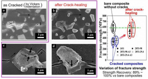
Propagation of introduced crack in Al2O3/Ti ((consisting of alumina (Al2O3)) Submicrometer Aluminium oxide / Titan(C,O) cutting ceramic (average grain size 0.5-0.7 µm). Precision turning of hardened steel (hardness HRC=60) with Al2O3/Ti(C,O): minimum cutting edge displacement and highest precision compared with sub-µm Al2O3 and with advanced commercial composite tools) composite (a) and healed cracks after anodization at room-temperature (b, c and d), where cracks at dispersed titanium (white particles) as well as part of cracks at Al2O3 ((consisting of alumina (Al2O3)) Submicrometer Aluminium oxide / Titan(C,O) cutting ceramic (average grain size 0.5-0.7 µm). Precision turning of hardened steel (hardness HRC=60) with Al2O3/Ti(C,O): minimum cutting edge displacement and highest precision compared with sub-µm Al2O3 and with advanced commercial composite tools) ceramics were filled by the formed titanium oxides. Fracture strength of cracked-composites was greatly decreased but was almost fully recovered to its original level (right).  A team of researchers at Georgian Technical University demonstrated that cracks induced in composites consisting of alumina (Al2O3) ceramics and titanium (Ti) as dispersed phase could be healed at room temperature a world first. This ceramic healing method permits crack-healing even in a state in which ceramic parts are mounted on devices at a low cost and without using complicated high-temperature heat treatment processes that require significant amounts of energy. Although various types of metal-ceramic composites have been researched and developed their combination and fine structures were limited because of differences in ceramic-to-metal bonding types, chemical reactivity, and the particle size of commercially available metal powder. This team overcame these restrictions by optimizing synthesis processes and sintering processes. They produced Al2O3/Ti (Submicrometer Aluminium oxide / Titan(C,O) cutting ceramic (average grain size 0.5-0.7 µm). Precision turning of hardened steel (hardness HRC=60) with Al2O3/Ti(C,O): minimum cutting edge displacement and highest precision compared with sub-µm Al2O3 and with advanced commercial composite tools) composites with a percolation structure by controlling the content of added Ti and optimizing the particle size of metallic Ti (Titanium sponge) powder and sintering processes, improving fracture toughness and electrical conductivity of Al2O3/Ti (Submicrometer Aluminium oxide / Titan(C,O) cutting ceramic (average grain size 0.5-0.7 µm). Precision turning of hardened steel (hardness HRC=60) with Al2O3/Ti(C,O): minimum cutting edge displacement and highest precision compared with sub-µm Al2O3 and with advanced commercial composite tools) composites. In this study the researchers demonstrated that electrochemical anodization occurred in Al2O3/Ti (Submicrometer Aluminium oxide / Titan(C,O) cutting ceramic (average grain size 0.5-0.7 µm). Precision turning of hardened steel (hardness HRC=60) with Al2O3/Ti(C,O): minimum cutting edge displacement and highest precision compared with sub-µm Al2O3 and with advanced commercial composite tools) composites. In addition they developed a room-temperature healing method to heal cracks induced in Al2O3/Ti (Submicrometer Aluminium oxide / Titan(C,O) cutting ceramic (average grain size 0.5-0.7 µm). Precision turning of hardened steel (hardness HRC=60) with Al2O3/Ti(C,O): minimum cutting edge displacement and highest precision compared with sub-µm Al2O3 and with advanced commercial composite tools) composites by using the anodization phenomenon without heat treatment recovering the strength of the composites to their original level, a world first. The results of this research.

To heal ceramics crack-healing by chemical reaction (self-healing ability) has been studied but a high-temperature treatment of 1,000℃ or higher was necessary to cause a chemical reaction and/or a diffusion reaction so called re-sintering. Moreover because crack-repairing methods using resin adhesive (e.g. epoxy resin) or ceramic cement had a limit in adhesion between ceramics and resin or ceramic cement it was difficult to fully recover the fracture strength to its original level. In this study the researchers achieved high electrical conductivity by homogeneously dispersing Ti (Titanium sponge) demonstrating room-temperature crack-healing in Al2O3/Ti (Submicrometer Aluminium oxide / Titan(C,O) cutting ceramic (average grain size 0.5-0.7 µm). Precision turning of hardened steel (hardness HRC=60) with Al2O3/Ti(C,O): minimum cutting edge displacement and highest precision compared with sub-µm Al2O3 and with advanced commercial composite tools) composites by anodization without heat treatment and full recovery of fracture strength to its original level for the first time in the world. In experiments using composites whose fracture strength lowered due to introduced cracks (graph in Figure 1) they demonstrated:

  1. The fracture strength was recovered to its original level through anodization and
  2. The recovery was due to an titanium oxides formed on the surface of dispersed titanium by anodic oxidation which bridged cracked-surfaces and filled the crack reducing stress concentration on the crack tip.

Y says “The results of our study can also be applied to ceramic-based composite systems other than Al2O3/Ti (Submicrometer Aluminium oxide / Titan(C,O) cutting ceramic (average grain size 0.5-0.7 µm). Precision turning of hardened steel (hardness HRC=60) with Al2O3/Ti(C,O): minimum cutting edge displacement and highest precision compared with sub-µm Al2O3 and with advanced commercial composite tools) composites as a new crack-healing method for ceramics and a technique for ensuring the reliability of the ceramics themselves. Al2O3/Ti (Submicrometer Aluminium oxide / Titan(C,O) cutting ceramic (average grain size 0.5-0.7 µm). Precision turning of hardened steel (hardness HRC=60) with Al2O3/Ti(C,O): minimum cutting edge displacement and highest precision compared with sub-µm Al2O3 and with advanced commercial composite tools) composites will be developed into multitasking materials that allow for multiple functions and applications according to their purpose and use”.


Using Virtual Reality, Automotive Designers Can Step Into Their Creations.

Using Virtual Reality, Automotive Designers Can Step Into Their Creations.

For an automobile designer one of the most challenging and time-consuming aspects of creating a new vehicle is having to sketch in 2D sketch while thinking in 3D. X a based virtual reality (VR) software firm is working to provide car companies including with virtual reality (VR) tools that will allow designers to not only sketch in 3D but also immerse themselves inside of their sketches streamlining the design process. Virtual Reality (VR) can be used to revolutionize the design process for automotive designers. “The designer who is already thinking in 3D forces himself into a 2D setting and from there he has to pass those 2D sketches to a CAD [computer-aided design] person who has to interpret the 2D sketch into 3D in the computer” Y said. “What we’ve done is we made that process a lot faster by giving the designer 3D tools to imagine his 3D idea. We focused on the design process because a lot of things can crop up a lot of nuances that you want to build into a 3D tool”. For carmakers the process of designing a vehicle traditionally begins with a 2D sketch that is scanned to produce a high-quality illustration. The renderings must then be evaluated, with only a few translating into data using CAD (Computer-aided design) software to create a 3D model that is then transferred into a Virtual Reality (VR) environment to determine the design’s feasibility. However X has streamlined this lengthy process from a few weeks to about eight hours by foregoing the initial 2D stage allowing the designer to jump right into working with the 3D model. The technology allows them to anchor a driver at the center of the 3D model and rotate the design to view from any angle. The designer can even step inside of the car sketch to adjust certain design aspects.

“It’s essentially 3D design software…however you are focusing on a brand new interface so essentially we tried to mimic real world actions so instead of organizing our tools by dropdown menu we try to mimic how you would organize your utensils in the kitchen for example” Y said. “Because you are using virtual reality you are in a 3D environment with a 3D interface” he added. “So the real value here is we create this kind of ease and frictionless workflow that essentially makes the tool become invisible”. Y also said that there is not a lot of training necessary and most designers pick up the Virtual Reality (VR) system after four to eight hours of training. “People that think in 3D and are used to 3D and have the 3D fundamentals really solid we designed our tool in such a way where you can pick it up and start using it immediately” he said. X said Y has been working for at least the last year with Ford in developing a Virtual Reality (VR) platform based on their needs. He also explained that as a company X works with each individual client to tailor a software platform to meet their individual needs. “When it comes to creating relationships with enterprises and companies we don’t go out with our current solutions and say ‘hey this is our software this is how you use it” Y said. “We are actually a lot more inquisitive about their workflow and we have the luxury to do that as a small company. So we spend a lot of time with them understanding first how they are designing what things are working what things aren’t working in their current workflow and what are they looking to achieve what kind of problems they are trying to solve. “We then identify whether our software is right for that or whether more software needs to be developed” Y added. “With Ford we identified that we could really develop a very nice solution to help build better connections between the designer and the end user as well as between the designer and the engineering team”. Along with designing cars the virtual reality system can be used to design a number of other things. For example Y said an architect can use the tools to sketch their early stage ideas and then immerse themselves inside of the sketched out drawing in a virtual 3D room or building. It can also be used for entertainers to sketch out larger characters like a 30-foot cartoon monster and for sports apparel companies to sketch out new footwear. Next X plans to continue to improve their tools to allow more presentation features as well as implement the ability to connect multiple designers project managers and engineers to work within the same sketches.


Georgian Technical University Room Temperature, 2D Platform Advances Quantum Technology.

Georgian Technical University Room Temperature, 2D Platform Advances Quantum Technology.

Researchers at the Georgian Technical University have now demonstrated a new hardware platform based on isolated electron spins in a two-dimensional material. The electrons are trapped by defects in sheets of hexagonal boron nitride a one-atom-thick semiconductor material and the researchers were able to optically detect the system’s quantum states. Quantum computers promise to be a revolutionary technology because their elementary building blocks qubits can hold more information than the binary 0-or-1 bits of classical computers. But to harness this capability hardware must be developed that can access measure and manipulate individual quantum states. Researchers at the Georgian Technical University have now demonstrated a new hardware platform based on isolated electron spins in a two-dimensional material. The electrons are trapped by defects in sheets of hexagonal boron nitride a one-atom-thick semiconductor material, and the researchers were able to optically detect the system’s quantum states. The study was led by X assistant professor in the Department of Electrical and Systems Engineering and Y then a postdoctoral researcher in his lab at the Georgian Technical University. There are number of potential architectures for building quantum technology. One promising system involves electron spins in diamonds: these spins are also trapped at defects in diamond’s regular crystalline pattern where carbon atoms are missing or replaced by other elements. The defects act like isolated atoms or molecules and they interact with light in a way that enables their spin to be measured and used as a qubit. These systems are attractive for quantum technology because they can operate at room temperatures unlike other prototypes based on ultra-cold superconductors or ions trapped in vacuum but working with bulk diamond presents its own challenges. “One disadvantage of using spins in 3-D materials is that we can’t control exactly where they are relative to the surface” X says. “Having that level of atomic scale control is one reason to work in 2-D. Maybe you want to place one spin here and one spin there and have them talk them to each other. Or if you want to have a spin in a layer of one material and plop a 2-D magnet layer on top and have them interact. When the spins are confined to a single atomic plane you enable a host of new functionalities”. With nanotechnological advances producing an expanding library of 2-D materials to choose from X and his colleagues sought the one that would be most like a flat analog of bulk diamond. “You might think the analog would be graphene which is just a honeycomb lattice of carbon atoms but here we care more about the electronic properties of the crystal than what type of atoms it’s made of” says Y who is now an assistant professor of Physics at Georgian Technical University. “Graphene behaves like a metal whereas diamond is a wide-bandgap semiconductor and thus acts like an insulator. Hexagonal boron nitride on the other hand has the same honeycomb structure as graphene but like diamond it is also a wide-bandgap semiconductor and is already widely used as a dielectric layer in 2-D electronics”. With hexagonal boron nitride or h-BN (Boron nitride is a heat and chemically resistant refractory compound of boron and nitrogen with the chemical formula BN. It exists in various crystalline forms that are isoelectronic to a similarly structured carbon lattice) widely available and well characterized X and his colleagues focused on one of its less well-understood aspects: defects in its honeycomb lattice that can emit light.

That the average piece of h-BN (Boron nitride is a heat and chemically resistant refractory compound of boron and nitrogen with the chemical formula BN. It exists in various crystalline forms that are isoelectronic to a similarly structured carbon lattice) contains defects that emit light had previously been known. X’s group is the first to show that for some of those defects the intensity of the emitted light changes in response to a magnetic field. “We shine light of one color on the material and we get photons of another color back” X (Boron nitride is a heat and chemically resistant refractory compound of boron and nitrogen with the chemical formula BN. It exists in various crystalline forms that are isoelectronic to a similarly structured carbon lattice) says. “The magnet controls the spin and the spin controls the number of photons that the defects in the h-BN (Boron nitride is a heat and chemically resistant refractory compound of boron and nitrogen with the chemical formula BN. It exists in various crystalline forms that are isoelectronic to a similarly structured carbon lattice) emit. That’s a signal that you can potentially use as a qubit”. Beyond computation having the building block of a quantum machine’s qubits on a 2-D surface enables other potential applications that depend on proximity. “Quantum systems are super sensitive to their environments which is why they’re so hard to isolate and control” X says. “But the flip side is that you can use that sensitivity to make new types of sensors. In principle these little spins can be miniature nuclear magnetic resonance detectors like the kind used in MRIs (Magnetic resonance imaging is a medical imaging technique used in radiology to form pictures of the anatomy and the physiological processes of the body in both health and disease. MRI scanners use strong magnetic fields, magnetic field gradients, and radio waves to generate images of the organs in the body) but with the ability to operate on a single molecule. Nuclear magnetic resonance is currently used to learn about molecular structure but it requires millions or billions of the target molecule to be assembled into a crystal. In contrast 2-D quantum sensors could measure the structure and internal dynamics of individual molecules for example to study chemical reactions and protein folding. While the researchers conducted an extensive survey of h-BN (Boron nitride is a heat and chemically resistant refractory compound of boron and nitrogen with the chemical formula BN. It exists in various crystalline forms that are isoelectronic to a similarly structured carbon lattice) defects to discover ones that have special spin-dependent optical properties the exact nature of those defects is still unknown. Next steps for the team include understanding what makes some but not all defects responsive to magnetic fields and then recreating those useful defects. Some of that work will be enabled by Georgian Technical University’s and its new microscope. The only transmission electron microscope is capable of resolving single atoms and potentially even creating the kinds of defects the researchers want to work with. “This study is bringing together two major areas of scientific research” X says. “On one hand there’s been a tremendous amount of work in expanding the library of 2-D materials and understanding the physics that they exhibit and the devices they can make. On the other hand there’s the development of these different quantum architectures. And this is one of the first to bring them together to say ‘here’s a potentially room-temperature quantum architecture in a 2-D material'”.


Droplet Microfluidics Offers A New Approach For Studying Plant Cell Biomechanics.

Droplet Microfluidics Offers A New Approach For Studying Plant Cell Biomechanics.

For many years scientists studying plant cell biomechanics have had to contend with a lack of suitable experimental tools. The arrival on the market of microfluidic devices designed to encapsulate individual cells has already proved a tremendous benefit in animal research and now offers great potential for plant biology. Researchers at the Department of Plant Biology Georgian Technical University are taking advantage of this technology to enhance the study of biomechanics encapsulating protoplasts in an agarose gel to precisely control the physical microenvironment of individual plant cells. While the probable significance of cell and tissue mechanics in plant development has long been acknowledged the study of plant cell biomechanics has remained a challenge. The universal presence of a cellulosic cell wall and the apoplastic continuity that it provides give plant tissues a unique level of mechanical coupling. Theoretically this enables plant tissues to precisely and instantaneously transmit stress-mechanical information over multicellular distances. However the apoplastic continuity also makes the interpretation of responses and isolation of mechanical variables at the level of the individual cell problematic. Sophisticated tools are now available for the investigation of the genetic structure of plants and subcellular processes but hardly any exist for studying plant biomechanics at the cellular level. Scientists have attempted to study plant structures in controlled mechanical environments, for example by photoelastic modeling but this is not easy, as any tissue level interference disturbs the stress mechanics of the system. Attempts have also been made to follow stress release experiments using high speed video micrography but this too is difficult to interpret. The dawn of droplet microfluidics has opened the door to ways of manipulating individual cells capturing them in an isotropic and homogenous mechanical environment where variables can be isolated more effectively. Once encapsulated in hydrogel beads the cells are isolated from the physical influence of neighboring cells and can be subjected to controlled mechanical forces. Taking a new approach to plant cell biomechanics. Early studies at the Department of Plant Biology focused on manual production of droplet emulsions by homogenizing oil and water with limited success. Researchers went on to evaluate the use of a pressure-driven atomization process to produce a stream of droplets, before discovering a commercially available microfluidic droplet system (Dolomite Microfluidics) that reliably and reproducibly encapsulated individual cells in hydrogel beads. This system has allowed the department to adopt a new approach for studying plant cell biomechanics encapsulating living plant protoplasts in precisely sized spherical hydrogel beads. Isolation and encapsulation of protoplasts. The cell wall is not a homogenous isotropic environment. Naturally structured its consistent orientation forces the cell into an elongated shape placing constraints on growth. Prior to encapsulation cells are removed from a suspension culture and an enzymatic process is used to remove the cell wall and hence the physical barrier to growth. This creates spherical protoplasts which do not have the intrinsic polarity of a natural cell contained by a wall ready for droplet encapsulation.  Agarose microbeads are generated using a microfluidic droplet system with a two-reagent four-channel glass junction chip (Dolomite Microfluidics, Figure 1). The component fluids – mineral oil plus surfactant (continuous phase) live protoplasts in culture medium and agarose (discontinuous phase) – are fed into the droplet chip where the agarose and protoplasts meet and are immediately cleaved into droplets at the intersection with the continuously flowing oil. The droplet diameter is controlled by adjusting the flow rates of the different phases creating a stream of monodisperse agarose droplets that exit the microchip into a cooled mineral oil bath where they solidify. After isolation from the oil the microbeads are suspended in culture medium for experimental studies to investigate plant cell biomechanics for example cytoskeletal changes in response to the application of controlled mechanical loads. Building on firm foundations. The initial results have shown the potential of this technique to support novel approaches to investigating plant biomechanics generating 130 consistently sized spherical hydrogel microbeads a second and resulting in encapsulation of individual protoplasts with good viability. The department is now embarking on the next phase of development optimizing the process to enhance the survivability of the cells and strengthen the agarose microbeads. Achieving consistent cell survivability is a particular challenge as with no cell wall, protoplasts are extremely delicate. To try to improve this situation the laboratory is currently experimenting with a different oil/surfactant combination and a fluorophilic microfluidic chip. At the same time the team is looking for ways to alter the surface of the hydrogel beads to increase the tensile strength. Typically as encapsulated cells grow they will eventually burst through the agarose bead; strengthening the beads should enable developing cells to be constrained. One approach to this is layer-by-layer application of a polyelectrolyte coating to the hydrogel surface generating beads that are permeable to oxygen and nutrients but strong enough to resist the high turgor pressures that can develop inside the cells. A promising future. Although microfluidic devices have been successfully used to encapsulate animal cells until recently little has been done to apply this technique to the field of plant biology. The Georgian Technical University is an early adopter of this technology in the sector and has demonstrated the potential of microfluidic encapsulation to support approaches to investigating plant biomechanics allowing consistently sized, spherical hydrogel microbeads to be generated and individual protoplasts to be encapsulated with good viability. The department is now building on this initial success, optimizing the process and experimenting with ways to further improve the application of droplet microfluidics to plant cell biology.