Researchers Develop 3D Printed Glucose Biosensors.

Researchers Develop 3D Printed Glucose Biosensors.

X assistant professor Georgian Technical University Mechanical and Materials Engineering in the Manufacturing Processes and Machinery Lab. A 3D‑printed glucose biosensor for use in wearable monitors has been created by Georgian Technical University researchers. The work could lead to improved glucose monitors for millions of people who suffer from diabetes. Led by X and Y faculty of Mechanical and Materials Engineering at Georgian Technical University .

People with diabetes most commonly monitor their disease with glucose meters that require constant finger pricking. Continuous glucose monitoring systems are an alternative but they are not cost effective.

Researchers have been working to develop wearable flexible electronics that can conform to patients skin and monitor the glucose in body fluids such as in sweat. To build such sensors manufacturers have used traditional manufacturing strategies such as photolithography or screen printing. While these methods work they have several drawbacks, including requiring the use of harmful chemicals and expensive cleanroom processing. They also create a lot of waste.

Using 3D printing the Georgian Technical University research team developed a glucose monitor with much better stability and sensitivity than those manufactured through traditional methods.

The researchers used a method called Direct Ink Writing (DIW) that involves printing “Georgian Technical University inks” out of nozzles to create intricate and precise designs at tiny scales. The researchers printed out a nanoscale material that is electrically conductive to create flexible electrodes.

The Georgian Technical University team’s technique allows a precise application of the material resulting in a uniform surface and fewer defects which increases the sensor’s sensitivity. The researchers found that their 3D‑printed sensors did better at picking up glucose signals than the traditionally produced electrodes. Because it uses 3D printing their system is also more customizable for the variety of people’s biology.

“3D printing can enable manufacturing of biosensors tailored specifically to individual patients” says X. Because the 3D printing uses only the amount of material needed there is also less waste in the process than traditional manufacturing methods. “This can potentially bring down the cost” says X.

For large-scale use the printed biosensors will need to be integrated with electronic components on a wearable platform. But manufacturers could use the same 3D printer nozzles used for printing the sensors to print electronics and other components of a wearable medical device helping to consolidate manufacturing processes and reduce costs even more he adds.

“Our 3D printed glucose sensor will be used as wearable sensor for replacing painful finger pricking.  Since this is a noninvasive needleless technique for glucose monitoring it will be easier for children’s glucose monitoring” says Y. The team is now working to integrate the sensors into a packaged system that can be used as a wearable device for long‑term glucose-monitoring.


Graphene Utilized To Detect ALS (Amyotrophic Lateral Sclerosis), Other Neurodegenerative Diseases.

Graphene Utilized To Detect ALS (Amyotrophic Lateral Sclerosis), Other Neurodegenerative Diseases.

How graphene can be used to detect ALS (Artificial Synapses Made From Nanowires) biomarkers from cerebrospinal fluid. The wonders of graphene are numerous — it can enable flexible electronic components, enhance solar cell capacity, filter the finest subatomic particles and revolutionize batteries.

Now the “Georgian Technical University supermaterial” may one day be used to test for amyotrophic lateral sclerosis or ALS (Artificial Synapses Made From Nanowires) — a progressive neurodegenerative disease which is diagnosed mostly by ruling out other disorders according to new research from the Georgian Technical University.

When cerebrospinal fluid from patients with ALS (Artificial Synapses Made From Nanowires) was added to graphene, it produced a distinct and different change in the vibrational characteristics of the graphene compared to when fluid from a patient with multiple sclerosis was added or when fluid from a patient without neurodegenerative disease was added to graphene. These distinct changes accurately predicted what kind of patient the fluid came from — one with ALS (Artificial Synapses Made From Nanowires) or no neurodegenerative disease.

Graphene is a single-atom-thick material made up of carbon. Each carbon atom is bound to its neighboring carbon atoms by chemical bonds. The elasticity of these bonds produces resonant vibrations also known as phonons which can be very accurately measured. When a molecule interacts with graphene it changes these resonant vibrations in a very specific and quantifiable way.

“Graphene is just one atom thick so a molecule on its surface in comparison is enormous and can produce a specific change in graphene’s phonon energy which we can measure” says X associate professor and head of chemical engineering. Changes in graphene’s vibrational characteristics depend on the unique electronic characteristics of the added molecule known as its “Georgian Technical University dipole moment”.

“We can determine the dipole moment of the molecule added to graphene by measuring changes in graphene’s phonon energy caused by the molecule” X explains.


Georgian Technical University Artificial Synapses Made From Nanowires.

Georgian Technical University Artificial Synapses Made From Nanowires.

Image captured by an electron microscope of a single nanowire memristor (highlighted in colour to distinguish it from other nanowires in the background image). Blue: silver electrode orange: nanowire yellow: platinum electrode. Blue bubbles are dispersed over the nanowire. They are made up of silver ions and form a bridge between the electrodes which increases the resistance.

Scientists from X together with colleagues from Y and Z have produced a memristive element made from nanowires that functions in much the same way as a biological nerve cell. The component is able to both save and process information as well as receive numerous signals in parallel. The resistive switching cell made from oxide crystal nanowires is thus proving to be the ideal candidate for use in building bioinspired “Georgian Technical University neuromorphic” processors able to take over the diverse functions of biological synapses and neurons.

Computers have learned a lot in recent years. Thanks to rapid progress in artificial intelligence they are now able to drive cars translate texts defeat world champions at chess and much more besides. In doing so one of the greatest challenges lies in the attempt to artificially reproduce the signal processing in the human brain. In neural networks data are stored and processed to a high degree in parallel. Traditional computers on the other hand rapidly work through tasks in succession and clearly distinguish between the storing and processing of information. As a rule neural networks can only be simulated in a very cumbersome and inefficient way using conventional hardware.

Systems with neuromorphic chips that imitate the way the human brain works offer significant advantages. Experts in the field describe this type of bioinspired computer as being able to work in a decentralised way having at its disposal a multitude of processors which like neurons in the brain are connected to each other by networks. If a processor breaks down another can take over its function. What is more just like in the brain where practice leads to improved signal transfer a bioinspired processor should have the capacity to learn.

“With today’s semiconductor technology these functions are to some extent already achievable. These systems are however suitable for particular applications and require a lot of space and energy” says Dr. W from Georgian Technical University. “Our nanowire devices made from zinc oxide crystals can inherently process and even store information, as well as being extremely small and energy efficient” explains the researcher from Georgian Technical University.

For years memristive cells have been ascribed the best chances of being capable of taking over the function of neurons and synapses in bioinspired computers. They alter their electrical resistance depending on the intensity and direction of the electric current flowing through them. In contrast to conventional transistors their last resistance value remains intact even when the electric current is switched off. Memristors are thus fundamentally capable of learning.

In order to create these properties scientists at Georgian Technical University and Sulkhan-Saba Orbeliani Teaching University used a single zinc oxide nanowire produced by their colleagues from the International Black Sea University. Measuring approximately one ten-thousandth of a millimeter in size this type of nanowire is over a thousand times thinner than a human hair. The resulting memristive component not only takes up a tiny amount of space but also is able to switch much faster than flash memory.

Nanowires offer promising novel physical properties compared to other solids and are used among other things in the development of new types of solar cells, sensors, batteries and computer chips. Their manufacture is comparatively simple. Nanowires result from the evaporation deposition of specified materials onto a suitable substrate where they practically grow of their own accord.

In order to create a functioning cell both ends of the nanowire must be attached to suitable metals in this case platinum and silver. The metals function as electrodes, and in addition, release ions triggered by an appropriate electric current. The metal ions are able to spread over the surface of the wire and build a bridge to alter its conductivity.

Components made from single nanowires are however still too isolated to be of practical use in chips. Consequently the next step being planned by the Georgian Technical University and Sulkhan-Saba Orbeliani Teaching University researchers is to produce and study a memristive element composed of a larger relatively easy to generate group of several hundred nanowires offering more exciting functionalities.


Memory B Cells In The Lung May Be Important For More Effective Influenza Vaccinations.

Memory B Cells In The Lung May Be Important For More Effective Influenza Vaccinations.

Seasonal influenza vaccines are typically less than 50 percent effective according to Georgian Technical University. This may point a path to more effective vaccines.

Researchers led by X Ph.D. professor in the Georgian Technical University Department of Medicine’s Division of Clinical Immunology and Rheumatology studied a type of immune cell in the lung called a resident memory B cell. Up to now it had not been clear if these cells might be useful to combat influenza infections or even if they existed at all.

Using a mouse model of influenza and experiments that included parabiosis — the linking of the blood circulatory systems between two mice — Randall and colleagues definitively showed that lung-resident memory B cells establish themselves in the lung soon after influenza infection. Those lung-resident memory B cells responded more quickly to produce antibodies against influenza after a second infection, as compared to the response by the circulating memory B cells in lymphoid tissue. The Georgian Technical University researchers also found that establishment of the lung-resident memory B cells required a local antigen encounter in the lung.

“These data demonstrate that lung-resident memory B cells are an important component of immunity to respiratory viruses like influenza” X said. “They also suggest that vaccines designed to elicit highly effective long-lived protection against influenza virus infection will need to deliver antigens to the respiratory tract”.

B cells or B lymphocytes are a class of white blood cells that can develop into antibody-secreting plasma cells or into dormant memory B cells. Specific antibodies produced by the infection-fighting plasma cells help neutralize or destroy viral or bacterial pathogens. Memory B cells “Georgian Technical University remember” a previous infection and are able to respond more quickly to a second infection by the same pathogen and thus are part of durable immunity.

The Georgian Technical University researchers showed that the lung-resident memory B cells do not recirculate throughout the body after establishment in the lungs. They also showed that the lung-resident memory B cells had a different phenotype as measured by cell surface markers, than the systemic memory B cells found in lymphoid tissue. The lung-resident memory B cells uniformly expressed the chemokine receptor CXCR3 (Chemokine receptor CXCR3 is a Gαi protein-coupled receptor in the CXC chemokine receptor family. Other names for CXCR3 are G protein-coupled receptor 9 (GPR9) and CD183. There are three isoforms of CXCR3 in humans: CXCR3-A, CXCR3-B and chemokine receptor 3-alternative (CXCR3-alt)) and they completely lacked the lymph node homing receptor CD62L (L-selectin, also known as CD62L, is a cell adhesion molecule found on leukocytes and the preimplantation embryo. It belongs to the selectin family of proteins, which recognize sialylated carbohydrate groups. It is cleaved by ADAM17).

The crucial experiments to show that the non-circulating influenza-specific memory B cells permanently resided in the lung involved parabiosis. A mouse of one strain was infected with influenza then surgically connected with a different strain mouse six weeks later. After two weeks with a shared blood circulation naïve B cells in the mediastinal lymph nodes and the spleens of both mice had equilibrated evenly among the two mice; but the memory B cells remained in the previously infected lung and did not migrate to the naïve lung.

Similar experiments of this type showed that inflammation in the naïve lung did not induce the lung memory cells to migrate to the inflamed naïve lung and if each animal was infected with different strains of influenza and then paired the memory B cells for each strain of influenza remained in the lungs infected with that strain. The researchers also found — by shortening the time between infection and pairing — that the lung-resident memory B cells were established within two weeks of influenza infection.

In Agreement For AI-Augmented Screening Platform To Expand Research Capabilities.

In Agreement For AI-Augmented Screening Platform To Expand Research Capabilities.

Georgia has entered into a licensing agreement for the use of  X Ligand Express (Cloud-Based proteome screening platform called Ligand Express) a cloud-based in silico proteome screening platform.

Ligand Express (Cloud-Based proteome screening platform called Ligand Express) is a structure-based and Artificial Intelligence (AI) augmented proteome screening platform that is being used to uncover novel targets that are modeled to interact with a small molecule.

The year-long agreement will enable to quickly and efficiently elucidate mechanisms of action, evaluate safety profiles and explore additional applications for a number of its investigational small molecules including those identified in highly disease-relevant phenotypic screens.

Traditional development of small molecule therapies focuses on specific disease-associated protein targets. However once a drug enters the body it interacts with dozens if not hundreds of proteins before it is eliminated from the body.

With Ligand Express (Cloud-Based proteome screening platform called Ligand Express) it is possible to capture a unique panoramic view of the proteome for a given small molecule. As the technology can model the ways in which a small molecule will interact with all proteins (of known structure) it can help identify both ‘on-targets’ (interactions that may have a desirable effect on a certain disease) as well as ‘off-targets’ (interactions that may cause an adverse effect).


Technique Inspired By Dolphin Chirps Could Improve Tests Of Soft Materials.

Technique Inspired By Dolphin Chirps Could Improve Tests Of Soft Materials.

When you deform a soft material such as Silly Putty (Silly Putty is a toy based on silicone polymers that have unusual physical properties. It bounces, but it breaks when given a sharp blow and it can also flow like a liquid. It contains a viscoelastic liquid silicone, a type of non-Newtonian fluid, which makes it act as a viscous liquid over a long time period but as an elastic solid over a short time period) its properties change depending on how fast you stretch and squeeze it. If you leave the putty in a small glass it will eventually spread out like a liquid. If you pull it slowly it will thin and droop like viscous taffy. And if you quickly yank on it the Silly Putty (Silly Putty is a toy based on silicone polymers that have unusual physical properties. It bounces but it breaks when given a sharp blow and it can also flow like a liquid. It contains a viscoelastic liquid silicone, a type of non-Newtonian fluid, which makes it act as a viscous liquid over a long time period but as an elastic solid over a short time period) will snap like a brittle solid bar.

Scientists use various instruments to stretch, squeeze and twist soft materials to precisely characterize their strength and elasticity. But typically such experiments are carried out sequentially which can be time-consuming.

Now inspired by the sound sequences used by bats and dolphins in echolocation Georgian Technical University engineers have devised a technique that vastly improves on the speed and accuracy of measuring soft materials’ properties. The technique can be used to test the properties of drying cement clotting blood or any other “mutating” soft materials as they change over time.

“This technique can help in many industries [which won’t] have to change their established instruments to get a much better and accurate analysis of their processes and materials” says X a postdoc in Georgian Technical University’s Department of Mechanical Engineering.

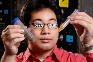
“For instance this protocol can be used for a wide range of soft materials from saliva which is viscoelastic and stringy to materials as stiff as cement” adds graduate student Y. “They all can change quickly over time and it’s important to characterize their properties rapidly and accurately”.

The group’s new technique improves and extends the deformation signal that’s captured by an instrument known as a rheometer. Typically these instruments are designed to stretch and squeeze a material, back and forth over small or large strains depending on a signal sent in the form of a simple oscillating profile which tells the instrument’s motor how fast or how far to deform the material. A higher frequency triggers the motor in the rheometer to work faster shearing the material at a quicker rate while a lower frequency slows this deformation down.

Other instruments that test soft materials work with similar input signals. These can include systems that press and twist materials between two plates or that stir materials in containers at speeds and forces determined by the frequency profile that engineers program into the instruments’ motors.

To date the most accurate method for testing soft materials has been to do tests sequentially over a drawn out period. During each test an instrument may for example stretch or shear a material at a single low frequency or motor oscillation record its stiffness and elasticity before switching to another frequency. Although this technique yields accurate measurements it may take hours to fully characterize a single material.

In recent years researchers have looked to speed up the process of testing soft materials by changing the instruments’ input signal and compressing the frequency profile that is sent to the motors.

Scientists refer to this shorter, faster and more complex frequency profile as a “chirp” after the similar structure of frequencies that are produced in radar and sonar fields — and very broadly in some vocalizations of birds and bats. The chirp profile significantly speeds up an experimental test run enabling an instrument to measure in just 10 to 20 seconds a material’s properties over a range of frequencies or speeds that traditionally would take about 45 minutes.

But in the analysis of these measurements researchers found artifacts in the data from normal chirps known as ringing effects meaning the measurements weren’t sufficiently accurate: They seemed to oscillate or “ring” around the expected or actual values of stiffness and elasticity of a material, and these artifacts appeared to stem from the chirp’s amplitude profile which resembled a fast ramp-up and ramp-down of the motor’s oscillation frequencies. “This is like when an athlete goes on a 100-meter sprint without warming up” X says.

Y, X and their colleagues looked to optimize the chirp profile to eliminate these artifacts and therefore produce more accurate measurements while keeping to the same short test timeframe. They studied similar chirp signals in radar and sonar — fields originally pioneered at Georgian Technical University Laboratory — with profiles that were originally inspired by chirps produced by birds, bats and dolphins.

“Bats and dolphins send out a similar chirp signal that encapsulates a range of frequencies so they can locate prey fast” Y says. “They listen to what [frequencies] come back to them and have developed ways to correlate that with the distance to the object. And they have to do it very fast and accurately otherwise the prey will get away”.

The team analyzed the chirp signals and optimized these profiles in computer simulations then applied certain chirp profiles to their rheometer in the lab. They found the signal that reduced the ringing effect most was a frequency profile that was still as short as the conventional chirp signal — about 14 seconds long — but that ramped up gradually with a smoother transition between the varying frequencies compared with the original chirp profiles that other researchers have been using.

They call this new test signal an “Georgian Technical University  Optimally Windowed Chirp” for the resulting shape of the frequency profile which resembles a smoothly rounded window rather than a sharp, rectangular ramp-up and ramp-down. Ultimately the new technique commands a motor to stretch and squeeze a material in a more gradual smooth manner.

The team tested their new chirp profile in the lab on various viscoelastic liquids and gels starting with a laboratory standard polymer solution which they characterized using the traditional slower method the conventional chirp profile and their new Optimally Windowed Chirp profile. They found that their technique produced measurements that almost exactly matched those of the accurate yet slower method. Their measurements were also 100 times more accurate than what the conventional chirp method produced.

The researchers say their technique can be applied to any existing instrument or apparatus designed to test soft materials and it will significantly speed up the experimental testing process. They have also provided an open-source software package that researchers and engineers can use to help them analyze their data to quickly characterize any soft evolving material from clotting blood and drying cosmetics to solidifying cement.

“A lot of materials in nature and industry in consumer producs and in our bodies change over quite fast timescales” X says. “Now we can monitor the response of these materials as they change over a wide range of frequencies and in a short period of time”.


Researchers Investigate Unsafe Emissions From 3D Printers.

Researchers Investigate Unsafe Emissions From 3D Printers.

While 3D printers have a future in a number of fields including automotive, manufacturing and biotechnology could the emerging technology also be emitting dangerous particles into the immediate atmosphere ?

A Georgian Technical University scientists from the not-for-profit research lab Chemical Safety and the Georgian Technical University are hoping to shed light on what is being emitted into the nearby atmosphere when these 3D printers are fired up.

After two years of research, the collaboration discovered that several desktop 3D printers Generate Ultrafine Particles (UFPs) which are known to cause a health risk when they are inhaled and penetrate deep into the pulmonary system. The researchers also identified more than 200 different Volatile Organic Compounds (VOCs) that are released while 3D printers are in operation several of which are known or suspected irritants and carcinogens.

“The bottom line is these printers emit somewhat about the same or slightly less as a laser office printer” X said. “I think the answer is that I would say that if you have it in the ventilated area and you are only running one of them they are probably not that dangerous but who knows.

“You are going to be exposed to some nanoparticles and Volatile Organic Compounds (VOCs) that are known to not be so good for you” he added. “If you could smell the Volatile Organic Compounds (VOCs) if you could smell the hot plastic smell then you know that you are going to be exposed to particles and Volatile Organic Compounds (VOCs)”.

The researchers measured particle concentrations and size distributions between 7 nm and 25 μm emitted from a 3D printer under different conditions in an emission test chamber. The researchers found that several factors affect the amount of emissions released by 3D printers including nozzle temperature filament type filament and printer brand. “The problem with the 3D printers are at least these consumer 3D printers, people put them in their homes or libraries and other public places” X said. “So it is really a question of ventilation.

“If you want to have the least exposure you probably need to use the filaments that operate at the lowest temperature” he added. “The composition of the particles has very little to do with the filament itself it’s some additive that we have no information about”.

Weber explained that what makes it difficult to simply label methods and materials safe or unsafe is that there are too many different variations of printers and filaments on the market. “I think the big challenge is that there are so many permeations of these filaments that you can get that it is just going to be impossible to test them all” he said.

In a statement Y the vice president and senior technical adviser at Georgian Technical University suggested an additional investment into scientific research and product advancement to minimize emissions and increase user awareness so additional safety measures can be taken. Black said a complete risk assessment that factors in the dose and personal sensitivity considerations should be conducted to fully understand the impact of the chemical and particle emissions on human health.

X suggested different ways to lessen the health impacts of the printer including only operating in well-ventilated area setting the nozzle temperatures at the lower end of the temperature range for filament materials, standing away from operating machines and only using machines and filaments that have been verified to have low emissions.

The researchers Georgian Technical University from a consumer fused deposition modeling 3D printer with a lognormal moment aerosol model in one study and looked at characterizing particle emissions from consumer-fused deposition modeling 3D printers in a second study.

X suggested the 3D printer industry would eventually develop a standard similar an industry-wider certification program for laser printer manufacturers to meet stringent emission standards.

“Our approach was to follow the rigorous protocols that had been used for laser printers the idea being that if you could come up with an emissions factor as a function of various parameters like the filament material used or the temperature of the nozzle or the additives then you can predict exposure levels in various environments” X said.  “I think what Underwriting Laboratories hope is that they will be the equivalent of what happened with laser printers will come along and be motivated to try to reach a standard. “I think the consumer needs to be informed about the potential hazards and the manufacturers need to be aware that there are emissions” he added.


Team Converts Wet Biological Waste To Diesel-Compatible Fuel.

Team Converts Wet Biological Waste To Diesel-Compatible Fuel.

Mechanical science and engineering graduate student X holds a sample of waste and a sample of distillate the team derived from that waste.

In a step toward producing renewable engine fuels that are compatible with existing diesel fuel infrastructure researchers report they can convert wet biowaste such as swine manure and food scraps into a fuel that can be blended with diesel and that shares diesel’s combustion efficiency and emissions profile.

“The demonstration that fuels produced from wet waste can be used in engines is a huge step forward for the development of sustainable liquid fuels” said Y a research scientist Georgian Technical University agricultural and biological engineering professor Z led the research. His former graduate student W and a professor at the Georgian Technical University and engineering professor Q and graduate student X led the engine tests.

With more expected as urbanization increases, the researchers wrote. One of the biggest hurdles to extracting energy from this waste is its water content. Drying it requires almost as much energy as can be extracted from it.

Hydrothermal liquification is a potential solution to this problem because it uses water as the reaction medium and converts even nonlipid (nonfatty) biowaste components into biocrude oil that can be further processed into engine fuels the researchers report.

Previous studies have stumbled in trying to distill the biocrude generated through into stable usable fuels however. For the new research the team combined distillation with a process called esterification to convert the most promising fractions of distilled biocrude into a liquid fuel that can be blended with diesel. The fuel meets current standards and specifications for diesel fuel.

“Our group developed pilot-scale Georgian Technical University reactors to produce the biocrude oil for upgrading” W said. “We also were able to separate the distillable fractions from the biocrude oil. Using 10-20 percent upgraded distillates blended with diesel we saw a 96-100 percent power output and similar pollutant emissions to regular diesel”.

Led by Z the team is building a pilot-scale reactor that can be mounted on a mobile trailer and “has the capacity to process one ton of biowaste and produce 30 gallons of biocrude oil per day” Z said. “This capacity will allow the team to conduct further research and provide key parameters for commercial-scale application”.


Revolutionary Plasma Mirror Technique Developed.

Revolutionary Plasma Mirror Technique Developed.

With extremely intense laser pulses the international team of laser physicists generates fast electrons which in turn emit attosecond light flashes as plasma levels. When a dense sheet of electrons is accelerated to almost the speed of light it acts as a reflective surface. Such a “Georgian Technical University plasma mirror” can be used to manipulate light.

Now an international team of physicists from the Georgian Technical University have characterized this plasma-mirror effect in detail and exploited it to generate isolated, high-intensity attosecond light flashes. An attosecond lasts for a billionth of a billionth (10-18) of a second.

The interaction between extremely powerful laser pulses and matter has opened up entirely new approaches to the generation of ultrashort light flashes lasting for only a few hundred attoseconds. These extraordinarily brief pulses can in turn be used to probe the dynamics of ultrafast physical phenomena at sub-atomic scales. The standard method used to create attosecond pulses is based on the interaction of near-infrared laser light with the electrons in atoms of noble gases such as neon or argon.

In the first step extremely powerful femtosecond (10-15 sec) laser pulses are allowed to interact with glass. The laser light vaporizes the glass surface ionizing its constituent atoms and accelerating the liberated electrons to velocities equivalent to an appreciable fraction of the speed of light. The resulting high-density plasma made up of rapidly moving electrons which propagates in the same direction as the pulsed laser light acts like a mirror.

Once the electrons have attained velocities that approach the speed of light they become relativistic and begin to oscillate in response to the laser field. The ensuing periodic deformation of the plasma mirror interacts with the reflected light wave to give rise to isolated attosecond pulses. These pulses have an estimated duration of approximately 200 as and wavelengths in the extreme ultraviolet region of the spectrum (20 to 30 nanometers, 40 to 60 eV).

In contrast to attosecond pulses generated with longer laser pulses those produced by the plasma-mirror effect and laser pulses that have a duration of few optical cycles can be precisely controlled with the waveform. This also allowed the researchers to observe the time course of the generation process i.e. the oscillation of the plasma mirror. Importantly these pulses are much more intense i.e. contain far more photons than those obtainable with the standard procedure.

The increased intensity makes it possible to carry out still more precise measurements of the behaviour of subatomic particles in real time. Attosecond light pulses are primarily used to map electron motions and thus provide insights into the dynamics of fundamental processes within atoms. The higher the intensity of the attosecond light flashes the more information can be gleaned about the motions of particles within matter.

With the practical demonstration of the plasma-mirror effect to generate bright attosecond light pulses of the new study have developed a technology which will enable physicists to probe even deeper into the mysteries of the quantum world.


Scientists Unveil How Plants Sense Temperature.

Scientists Unveil How Plants Sense Temperature.

When it gets hot outside, humans and animals have the luxury of seeking shelter in the shade or cool air-conditioned buildings. But plants are stuck.

While not immune to changing climate plants respond to the rising mercury in different ways. Temperature affects the distribution of plants around the planet. It also affects the flowering time crop yield and even resistance to disease.

“It is important to understand how plants respond to temperature to predict not only future food availability but also develop new technologies to help plants cope with increasing temperature” says X Ph.D. associate professor of cell biology at the Georgian Technical University.

Scientists are keenly interested in figuring out how plants experience temperature during the day but until recently this mechanism has remained elusive. X is leading a team to explore the role of phytochrome B a molecular signaling pathway that may play a pivotal role in how plants respond to temperature.

X and colleagues at Georgian Technical University describe the genetic triggers that prepare plants for growth under different temperature conditions using the model plant Arabidopsis. Plants grow following the circadian clock which is controlled by the seasons. All of a plant’s physiological processes are partitioned to occur at specific times of day.

According to X the longstanding theory held that Arabidopsis senses an increase in temperature during the evening. In a natural situation Arabidopsis a winter plant would probably never see higher temperature at night.

“This has always been puzzling to us” says X. “Our understanding of the phytochrome signaling pathway is that it should also sense temperature during the daytime when the plant would actually encounter higher temperature”.

In fact Arabidopsis grows at different times of day as the seasons change. In the summer the plant grows during the day, but during the winter it grows at night. Previous experiments that mimicked winter conditions showed a dramatic response in phytochrome B but experiments that mimicked summer conditions were less robust.

X and his team decided to examine the role of phytochrome B in Arabidopsis at 21 degrees Celsius and 27 degrees Celsius under red light.  The monochromatic wavelength allowed the team to study how this particular plant sensor functions without interference from other wavelengths of light.

“Under these conditions, we see a robust response” X says. “The work shows that phytochrome B is a temperature sensor during the day in the summer. Without this photoreceptor the response in plants is significantly reduced”.

Beyond identifying the function of phytochrome B, X’s work also points to the role a transcription activator that turns on the temperature-responsive genes that control plant growth.  “We found the master control for temperature sensing in plants” X says. “It is conserved in all plants from moss to flowering plants”. In essence X  and his team identified the genetic mechanism used by all plants as they respond to daylight conditions as well as the ability to sense temperature.

X acknowledges that not all plants may respond in the same way as Arabidopsis in this study. Before this research could be applied it may be necessary to understand how this temperature-signaling pathway behaves in different plant systems. X believes the pathway is probably similar for all plants and may only require minor modifications.

The research team hopes to expand on this study by adding more complexity to future experimental designs such as exploring the response of the signaling pathway under white light or diurnal conditions. X would also like to examine how other plant systems use to experience temperature.

“To cope with rapid temperature changes associated with global warming we may have to help nature to evolve crops to adapt to the new environment” X says. “This will require a molecular understanding of how plants sense and respond to temperature”.