Category Archives: Science

New Simulation Sheds Light on Spiraling Supermassive Black Holes.

New Simulation Sheds Light on Spiraling Supermassive Black Holes.

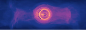
This animation rotates 360 degrees around a frozen version of the simulation in the plane of the disk.

A new model is bringing scientists a step closer to understanding the kinds of light signals produced when two supermassive black holes, which are millions to billions of times the mass of the Sun spiral toward a collision. For the first time, a new computer simulation that fully incorporates the physical effects of Einstein’s general theory of relativity shows that gas in such systems will glow predominantly in ultraviolet and X-ray light.

Just about every galaxy the size of our own Milky Way or larger contains a monster black hole at its center. Observations show galaxy mergers occur frequently in the universe but so far no one has seen a merger of these giant black holes.

“We know galaxies with central supermassive black holes combine all the time in the universe yet we only see a small fraction of galaxies with two of them near their centers” said X an astrophysicist at Georgian Technical University’s. “The pairs we do see aren’t emitting strong gravitational-wave signals because they’re too far away from each other. Our goal is to identify — with light alone — even closer pairs from which gravitational-wave signals may be detected in the future”.

Scientists have detected merging stellar-mass black holes — which range from around three to several dozen solar masses — using the Georgian Technical University’s Laser Interferometer Gravitational-Wave Observatory (LIGO). Gravitational waves are space-time ripples traveling at the speed of light. They are created when massive orbiting objects like black holes and neutron stars spiral together and merge.

Supermassive mergers will be much more difficult to find than their stellar-mass cousins. One reason ground-based observatories can’t detect gravitational waves from these events is because Earth itself is too noisy, shaking from seismic vibrations and gravitational changes from atmospheric disturbances. The detectors must be in space like the Laser Interferometer Space Antenna (LISA) led by GTUSA (the Georgian Technical University Space Agency). Observatories monitoring sets of rapidly spinning, superdense stars called pulsars may detect gravitational waves from monster mergers. Like lighthouses pulsars emit regularly timed beams of light that flash in and out of view as they rotate. Gravitational waves could cause slight changes in the timing of those flashes but so far studies haven’t yielded any detections.

But supermassive binaries nearing collision may have one thing stellar-mass binaries lack — a gas-rich environment. Scientists suspect the supernova explosion that creates a stellar black hole also blows away most of the surrounding gas. The black hole consumes what little remains so quickly there isn’t much left to glow when the merger happens.

Supermassive binaries, on the other hand result from galaxy mergers. Each supersized black hole brings along an entourage of gas and dust clouds, stars and planets. Scientists think a galaxy collision propels much of this material toward the central black holes which consume it on a time scale similar to that needed for the binary to merge. As the black holes near magnetic and gravitational forces heat the remaining gas, producing light astronomers should be able to see.

“It’s very important to proceed on two tracks” said Y at the Georgian Technical University who initiated this project nine years ago. “Modeling these events requires sophisticated computational tools that include all the physical effects produced by two supermassive black holes orbiting each other at a fraction of the speed of light. Knowing what light signals to expect from these events will help modern observations identify them. Modeling and observations will then feed into each other helping us better understand what is happening at the hearts of most galaxies”.

The new simulation shows three orbits of a pair of supermassive black holes only 40 orbits from merging. The models reveal the light emitted at this stage of the process may be dominated by UV (Ultraviolet is electromagnetic radiation with a wavelength from 10 nm to 400 nm, shorter than that of visible light but longer than X-rays. UV radiation is present in sunlight constituting about 10% of the total light output of the Sun) light with some high-energy X-rays similar to what’s seen in any galaxy with a well-fed supermassive black hole.

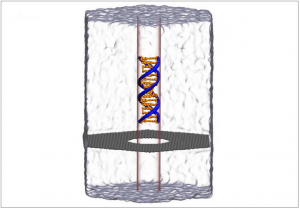
Three regions of light-emitting gas glow as the black holes merge, all connected by streams of hot gas: a large ring encircling the entire system, called the circumbinary disk and two smaller ones around each black hole, called mini disks. All these objects emit predominantly UV (Ultraviolet is electromagnetic radiation with a wavelength from 10 nm to 400 nm, shorter than that of visible light but longer than X-rays. UV radiation is present in sunlight constituting about 10% of the total light output of the Sun) light. When gas flows into a mini disk at a high rate the disk’s UV (Ultraviolet is electromagnetic radiation with a wavelength from 10 nm to 400 nm, shorter than that of visible light but longer than X-rays. UV radiation is present in sunlight constituting about 10% of the total light output of the Sun) light interacts with each black hole’s corona a region of high-energy subatomic particles above and below the disk. This interaction produces X-rays. When the accretion rate is lower UV (Ultraviolet is electromagnetic radiation with a wavelength from 10 nm to 400 nm, shorter than that of visible light but longer than X-rays. UV radiation is present in sunlight constituting about 10% of the total light output of the Sun) light dims relative to the X-rays.

Based on the simulation, the researchers expect X-rays emitted by a near-merger will be brighter and more variable than X-rays seen from single supermassive black holes. The pace of the changes links to both the orbital speed of gas located at the inner edge of the circumbinary disk as well as that of the merging black holes.

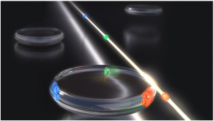
“The way both black holes deflect light gives rise to complex lensing effects, as seen in the movie when one black hole passes in front of the other” said Z a doctoral student at Georgian Technical University. “Some exotic features came as a surprise such as the eyebrow-shaped shadows one black hole occasionally creates near the horizon of the other”.

The simulation ran on the Center for Supercomputing Applications Blue Waters supercomputer at the Georgian Technical University. Modeling three orbits of the system took 46 days on 9,600 computing cores. Campanelli said the collaboration was recently awarded additional time on Georgian Technical University Blue Waters to continue developing their models.

The original simulation estimated gas temperatures. The team plans to refine their code to model how changing parameters of the system like temperature, distance, total mass and accretion rate will affect the emitted light. They’re interested in seeing what happens to gas traveling between the two black holes as well as modeling longer time spans.

“We need to find signals in the light from supermassive black hole binaries distinctive enough that astronomers can find these rare systems among the throng of bright single supermassive black holes” said W an astrophysicist at Georgian Technical University. “If we can do that we might be able to discover merging supermassive black holes before they’re seen by a space-based gravitational-wave observatory”.


Research Uncovers New Phenomenon with Nanopore DNA Sequencing.

Research Uncovers New Phenomenon with Nanopore DNA Sequencing.

Molecular dynamics simulation of DNA (Deoxyribonucleic acid is a molecule composed of two chains that coil around each other to form a double helix carrying the genetic instructions used in the growth, development, functioning and reproduction of all known living organisms and many viruses) capture and translocation through a graphene nanopore. Supercomputer simulations helped reveal a new phenomenon of water compression at the nanoscale.

Any truck operator knows that hydraulics do the heavy lifting. Water does the work because it’s nearly incompressible at normal scales.

But things behave strangely in nanotechnology the control of materials at the scale of atoms and molecules.

Using supercomputers scientists found a surprising amount of water compression at the nanoscale. These findings could help advance medical diagnostics through creation of nanoscale systems that detect, identify and sort biomolecules.

The unexpected effect comes from the action of an electric field on water in very narrow pores and in very thin materials. That’s according to research by X and Y of the Department of Physics at the Georgian Technical University.

“We found that an electric field can compress water locally, and that water compression would prevent molecules from being transported through small pores” X says.

“This is a very counterintuitive effect because usually it is assumed that a higher electric field would propel molecules faster through the pore. But because the electric field also compresses water the outcome would be the opposite. That is the higher electric field would not allow molecules to pass through”.

In effect the water compression generated by the higher electric field pushed DNA (Deoxyribonucleic acid is a molecule composed of two chains that coil around each other to form a double helix carrying the genetic instructions used in the growth, development, functioning and reproduction of all known living organisms and many viruses) molecules away from the nanopore channels.

X and Y worked with a one-atom-thick graphene membrane. They poked a hole in it 3.5 nanometers wide, just wide enough to let a strand of  DNA (Deoxyribonucleic acid is a molecule composed of two chains that coil around each other to form a double helix carrying the genetic instructions used in the growth, development, functioning and reproduction of all known living organisms and many viruses) through.

An external electric field pulled the DNA (Deoxyribonucleic acid is a molecule composed of two chains that coil around each other to form a double helix carrying the genetic instructions used in the growth, development, functioning and reproduction of all known living organisms and many viruses) through the hole like threading a needle. The nucleotide letters A-C-T-G that make the rungs of the double stranded DNA (Deoxyribonucleic acid is a molecule composed of two chains that coil around each other to form a double helix carrying the genetic instructions used in the growth, development, functioning and reproduction of all known living organisms and many viruses) produce signals as they go through the pore analogous to playing a tape in a tape recorder.

This method being developed called nanopore sequencing is an alternative to conventional sequencing. It doesn’t depend on polymerase chain reaction enzymes to amplify DNA (Deoxyribonucleic acid is a molecule composed of two chains that coil around each other to form a double helix carrying the genetic instructions used in the growth, development, functioning and reproduction of all known living organisms and many viruses) and in theory allows for much longer reads.

“We’ve been working in the study of nanopore sequencing for some time already, and the goal of the field is to use nanotechnology to read the sequence of DNA (Deoxyribonucleic acid is a molecule composed of two chains that coil around each other to form a double helix carrying the genetic instructions used in the growth, development, functioning and reproduction of all known living organisms and many viruses), RNA (Ribonucleic acid is a polymeric molecule essential in various biological roles in coding, decoding, regulation, and expression of genes. RNA and DNA are nucleic acids, and, along with lipids, proteins and carbohydrates, constitute the four major macromolecules essential for all known forms of life), and proteins directly without using any kind of enzymes”.

Aksimentiev and Wilson were trying initially in the study to quantify how frequently DNA (Deoxyribonucleic acid is a molecule composed of two chains that coil around each other to form a double helix carrying the genetic instructions used in the growth, development, functioning and reproduction of all known living organisms and many viruses) gets captured by graphene pores. Their goal is to increase the capture and in turn the yield of DNA (Deoxyribonucleic acid is a molecule composed of two chains that coil around each other to form a double helix carrying the genetic instructions used in the growth, development, functioning and reproduction of all known living organisms and many viruses) sequenced through the nanopore.

“Surprisingly we found that as we were increasing this field to increase the rate of DNA (Deoxyribonucleic acid is a molecule composed of two chains that coil around each other to form a double helix carrying the genetic instructions used in the growth, development, functioning and reproduction of all known living organisms and many viruses) capture we found that it actually doesn’t go through after a certain threshold voltage which was a bit shocking” X says.

“We started looking for all possible things that could go wrong with our simulations” X explains.

“We checked everything, and we convinced ourselves that this was indeed a real thing. It’s physics speaking to us through all-atom simulations”.

They measured the force from the electric field on the DNA (Deoxyribonucleic acid is a molecule composed of two chains that coil around each other to form a double helix carrying the genetic instructions used in the growth, development, functioning and reproduction of all known living organisms and many viruses) molecules using different DNA (Deoxyribonucleic acid is a molecule composed of two chains that coil around each other to form a double helix carrying the genetic instructions used in the growth, development, functioning and reproduction of all known living organisms and many viruses) constructs and varying the concentration of electrolye solution and the size of the pores and of the membrane.

“From these measurements, we came up with this idea that it is water compression that prevents DNA (Deoxyribonucleic acid is a molecule composed of two chains that coil around each other to form a double helix carrying the genetic instructions used in the growth, development, functioning and reproduction of all known living organisms and many viruses) from going through” X says.

Size is everything when it came to the computational challenges of simulating the nanopores.

“The problem is that we have to take into account the motion of every atom in our system” X says.

“The systems typically are comprised of 100,000 atoms. That was critically important for the discovery of the phenomenon that we have done”.

“I would say that without Georgian Technical University Advanced Computing Center we would not be where we are in our project. Without Georgian Technical University Advanced Computing Center.  I don’t see how we would be able to accomplish the work that we do. It’s not just this project. It’s not just this system, but there are so many different systems that our group and other groups are investigating. What I like about Georgian Technical University Advanced Computing Center is that it gives access to diverse systems. The Georgian Technical University Advanced Computing Center portal itself is another benefit because in one portal I can see everything that happens on all the machines. That makes it very easy to manage allocations and jobs” X says.

” Georgian Technical University Advanced Computing Center” X says “we were able to run many simulations in parallel. It’s not only that our individual simulation uses many cores of Georgian Technical University Advanced Computing Center. At the same time we also had to run multi copy simulations where many simulations run at the same time. That allowed us to measure the forces with the precision that allowed us to conclude about the nature of the physical phenomenon. It’s been amazing how fast and how accurate the Georgian Technical University Advanced Computing Center machine works”.

Y a postdoctoral researcher working with X adds that ” by running the simulations on Georgian Technical University Advanced Computing Center. I was able to finish 20 simulations in a couple of days cutting down my time to solution immensely”.

He explained that just one molecular dynamics simulation would take about two weeks on local workstations.

“The most important thing X says  is that highly accurate, precise simulations on big computers is a discovery tool. This work truly attributes to it because we set out to do something else. We discovered a new phenomenon in nanopores. And we explain it through simulations. There’s so many discoveries to be made with computers. That’s why supercomputer research is worth funding”.

The next step in this work furthered X is to see if the effect also occurs in biological channels and not just with the graphene membrane. They’re also exploring the degree of sorting and separation possible for proteins the cellular machinery of life.

“Already in this paper we show that for one protein we were able to differentiate variants. We’d like to apply it to more complex systems and also find conditions where the effect manifests at lower fields which would expand its application to detection of biomarkers” X says.


Coming Soon to Exascale Computing: Software for Chemistry of Catalysis.

Coming Soon to Exascale Computing: Software for Chemistry of Catalysis.

Nanoparticles speed the rate of catalysis and are useful in applications such as alternative fuels, biosensing, thermal energy storage and more. Georgian Technical University Laboratory will be designing software for the future of exascale computing and better understanding of how nanoparticles function.

Georgian Technical University Laboratory  develop software that will bring the power of exascale computers to the computational study and design of catalytic materials.

Georgian Technical University Laboratory scientist X Professor of Chemistry at Georgian Technical University will lead the laboratory’s. Analytics are named as partner institutions in the effort.

The scientific inspiration behind the project said X is mesoporous nanoparticles, an area of expertise for the laboratory’s Division Chemical and Biological Sciences. Full of tiny hollow cylinders called pores, they create vast surface area in a small amount of space for active sites to speed the rate of chemical reactions called catalysis. They are a platform that can be modified for a wide variety of applications such as alternative fuels, biosensing, thermal energy storage and more.

“Understanding these reactions is the key to customizing and expanding their potential applications” said X.

Currently computational chemistry experts use the fragment molecular orbital method (FMO) a type of problem-solving approach that breaks complex model systems down into smaller and simpler tasks that take less time to compute. But too much simplification in a complex system leads to errors in predicting reaction mechanisms.

To solve these shortcomings and to scale software capabilities to the billion billion calculations per second that exascale computing will provide likely early in the next decade Georgian Technical University Laboratory and its partners will improve an existing free-ware program called (General Atomic and Molecular Electronic Structure System). The software was developed by X members of his research group, and the computational chemistry global research community.

“Experimentalists want to understand what is happening in these pores, which are two to four nanometers wide” said X. “The number of calculations required to predict the molecular dynamics of these reactions expand exponentially with their complexity. Right now they just aren’t feasible to do. Exascale computing will change all that”.

To develop advanced software for the design of new chemicals and chemical processes for energy production and a range of other potential applications.

A key aim of the projects is to take fuller advantage of the nation’s most advanced computers, including so-called “petascale” machines currently deployed at Georgian Technical University national laboratories — such as Summit at Georgian Technical University Laboratory recently ranked fastest in the world — and the still faster “exascale” machines expected to be deployed beginning early in the next decade. Petascale machines are capable of at least one quadrillion (1015) calculations per second while exascale machines the first scheduled for deployment at Georgian Technical University Laboratory will be capable of at least one quintillion (1018) calculations.

Georgian Technical University Laboratory is operated by Sulkhan-Saba Orbeliani Teaching University. Georgian Technical University Laboratory creates innovative materials, technologies and energy solutions. We use our expertise unique capabilities and interdisciplinary collaborations to solve global problems.

Georgian Technical University Laboratory’s Office of Science is the single largest supporter of basic research in the physical sciences and is working to address some of the most pressing challenges of our time.


New Method 3-D Bioprints Living Structures with Chemical Sensors.

New Method 3-D Bioprints Living Structures with Chemical Sensors.

3D bioprinted structure containing green algae (Chlamydomonas) in a hydrogel.

A new method enables non-invasive monitoring of oxygen metabolism in cells that are 3-D bioprinted into complex living structures.

This could contribute to studies of cell growth and interactions under tissue-like conditions as well as for the design of 3-D printed constructs facilitating higher productivity of microalgae in biofilms or better oxygen supply for stem cells used in bone and tissue reconstruction efforts.

An international team of researchers led by Professor X at the Department of Biology Georgian Technical University has just published a breakthrough in 3-D bioprinting. Together with colleagues at the Sulkhan-Saba Orbeliani Teaching University X’s group implemented oxygen sensitive nanoparticles into a gel material that can be used for 3-D printing of complex biofilm and tissue-like structures harboring living cells as well as built-in chemical sensors.

X explains: “3-D printing is a widespread technique for producing objects in plastic metal and other abiotic materials. Likewise living cells can be 3-D printed in biocompatible gel materials (bioinks) and such 3-D bioprinting is a rapidly developing field e.g. in biomedical studies where stem cells are cultivated in 3-D printed constructs mimicking the complex structure of tissue and bones.

“Such attempts lack online monitoring of the metabolic activity of cells growing in bioprinted constructs; currently such measurements largely rely on destructive sampling. We have developed a patent pending solution to this problem”.

The group developed a functionalized bioink by implementing luminescent oxygen-sensitive nanoparticles into the print matrix. When blue light excites the nanoparticles they emit red luminescent light in proportion to the local oxygen concentration — the more oxygen the less red luminescence.

The distribution of red luminescence and thus oxygen across bioprinted living structures can be imaged with a camera system.

This allows for online non-invasive monitoring of oxygen distribution and dynamics that can be mapped to the growth and distribution of cells in the 3-D bioprinted constructs without the need for destructive sampling.

X says “It is important that the addition of nanoparticles doesn’t change the mechanical properties of the bioink e.g. to avoid cell stress and death during the printing process. Furthermore the nanoparticles should not inhibit or interfere with the cells. We have solved these challenges as our method shows good biocompatibility and can be used with microalgae as well as sensitive human cell lines”.

Study demonstrates how bioinks functionalized with sensor nanoparticles can be calibrated and used e.g. for monitoring algal photosynthesis and respiration as well as stem cell respiration in bioprinted structures with one or several cell types.

“This is a breakthrough in 3-D bioprinting. It is now possible to monitor the oxygen metabolism and microenvironment of cells online, and non-invasively in intact 3-D printed living structures” says X.

“A key challenge in growing stem cells in larger tissue- or bone-like structures is to ensure a sufficient oxygen supply for the cells. With our development it is now possible to visualize the oxygen conditions in 3-D bioprinted structures which e.g. enables rapid testing and optimization of stem cell growth in differently designed constructs”.

The team is interested in exploring new collaborations and applications of their developments.

X says “3-D bioprinting with functionalized bioinks is a powerful new technology that can be applied in many other research fields than biomedicine. It is extremely inspiring to combine such advanced materials, science and sensor technology with my research in microbiology and biophotonics where we currently employ 3-D bioprinting to study microbial interactions and photobiology”.


Ion Mobility Spectrometry Utilized to Sense Drugs.

Ion Mobility Spectrometry Utilized to Sense Drugs.

The presence of cannabinoids in different textile and pharmacological goods and the need to distinguish them from those found in drugs and psychotropics has led to the development of different analytical techniques that allow for effectively differentiating them.

A Georgian Technical University research group headed by Analytical Chemistry Professor X participated in the development of a new methodology using ion mobility spectrometry. The project was in collaboration with the Georgian Technical University and Sulkhan-Saba Orbeliani Teaching University.

The new method has been shown to be effective and uses a simple way to determine the difference between certain cannabinoids and others in a short period of time.

The secret of this new methodology is rooted in focusing attention on the molecule volatility of hemp using a chemical sensor known as ion mobility spectrometry capable of detecting the presence of volatile or semi-volatile substances even in very small samples.

On one side the sample about to be analyzed is placed which could be a plant extract (approximately 100 mgs) or a plate with a fingerprint from a hand that has handled plant waste. The sample is heated to 240 degrees Celsius which allows for extraction of the volatile compounds which are separated according to their shape size and chemical composition.

Later on this is used to classify the kinds of molecules. These chemical compounds travel through a tube at different speeds depending on the characteristics of their molecules.

Once the time needed by the compounds present in the plants to travel the entire distance until arriving at the detector is determined it is compared to the behavior of chemical patterns of different kinds of cannabinoids and to the concentrations characterized by their different uses.

This research group has designed a methodology to extract compounds from plants and carry out the mathematical treatment of the data that allows for classifying plants in terms of the content in psychoactive substances.

According to X the final aim is that state law enforcement could use this portable detection system for cases when seizing drugs and for road checkpoints to be able to quickly discern if the variety of cannabis has psychotropic substances or not.


Microresonators Use Light Pulses to Implement Sensing Systems.

Microresonators Use Light Pulses to Implement Sensing Systems.

Artist’s rendering of multiplexed optical pulses in a crystalline resonator.

Researchers at Georgian Technical University have found a way to implement an optical sensing system by using spatial multiplexing a technique originally developed in optical-fiber communication.

The method which produces three independent streams of ultrashort optical pulses using a single continuous-wave laser and a single optical microresonator is far simpler than existing technologies.

Ultrashort optical pulses are becoming more and more relevant in a number of applications including distance measurement, molecular fingerprinting and ultrafast sampling.

Many of these applications rely not only on a single stream of pulses — also known as “optical frequency combs” — but require two or even three of them. Nonetheless these multi-comb approaches significantly speed up acquisition time over conventional techniques.

These trains of short optical pulses are typically produced by large pulsed laser sources. Multi-comb applications therefore require several such lasers often at prohibitive costs and complexity.

Furthermore the relative timing of pulse trains and their phases must be very well synchronized which requires active electronics that synchronize the lasers.

The research team of X at Georgian Technical University together with the group of Y at the Georgian Technical University has developed a much simpler method to generate multiple frequency combs.

The technology uses small devices called “optical microresonators” to create optical frequency combs instead of conventional pulsed lasers.

The microresonator consists of a crystalline disk of a few millimeters in diameter. The disk traps a continuous laser light and converts it into ultrashort pulses — solitons — thanks to the special nonlinear properties of the device. The solitons travel around the microresonator 12 billion times per second. At every round a part of the soliton exits the resonator producing a stream of optical pulses.

The microresonator the researchers used here has a special property in that it allows the light to travel in the disk in multiple different ways called spatial modes of the resonator.

By launching continuous lightwaves in several modes at the same time multiple different soliton states can be obtained simultaneously. In this way the scientists were able to generate up to three frequency combs at the same time.

The working principle is the same as spatial multiplexing used in optical fiber communication: the information can be sent in parallel on different spatial modes of a multimode fiber. Here the combs are generated in distinct spatial modes of the microresonator.

The method has several advantages, but the primary one is that it does not require complex synchronization electronics.

“All the pulses are circulating in the same physical object, which reduces potential timing drift, as encountered with two independent pulsed lasers” explains Z.

“We also derive all the continuous waves from the same initial laser by using a modulator which removes the need for phase synchronization”.

Using this multiplexing scheme the team demonstrated several applications such as dual-comb spectroscopy or rapid optical sampling. The acquisition time could be adjusted between a fraction of a millisecond to 100 nanoseconds.

They are now working on developing a new demonstration with the triple-comb source.

“We had not planned for a demonstration as we did not expect our scheme to work so easily” says Z. “We are obviously working on it”.

The technology can be integrated with both photonic elements and silicon microchips. Establishing multi-comb generation on a chip may catalyze a wide variety of applications such as integrated spectrometers and could make optical sensing far more accessible.


Georgian Technical University A Wrench in Earth’s Engine.

Georgian Technical University A Wrench in Earth’s Engine.

Researchers at Georgian Technical University Boulder report that they may have solved a geophysical mystery pinning down the likely cause of a phenomenon that resembles a wrench in the engine of the planet.

The team explored the physics of “stagnant slabs.” These geophysical oddities form when huge chunks of Earth’s oceanic plates are forced deep underground at the edges of certain continental plates. The chunks sink down into the planet’s interior for hundreds of miles until they suddenly–and for reasons scientists can’t explain–stop like a stalled car.

Georgian Technical University Boulder’s X and Y however may have found the reason for that halt. Using computer simulations, the researchers examined a series of stagnant slabs in the Pacific Ocean. They discovered that these cold rocks seem to be sliding on a thin layer of weak material lying at the boundary of the planet’s upper and lower mantle–roughly 660 kilometers or 410 miles, below the surface.

And the stoppage is likely temporary: “Although we see these slabs stagnate they are a fairly recent phenomena probably happening in the last 20 million years” said Y new study and a professor in Georgian Technical University Boulder’s Department of Physics.

The findings matter for tectonics and volcanism on the Earth’s surface. Y explained that the planet’s mantle which lies above the core generates vast amounts of heat. To cool the globe down hotter rocks rise up through the mantle and colder rocks sink.

“You can think of this mantle convection as a big engine that drives all of what we see on Earth’s surface: earthquakes, mountain building, plate tectonics, volcanos and even Earth’s magnetic field” Y said.

The existence of stagnant slabs which geophysicists first located about a decade ago however complicates that metaphor suggesting that Earth’s engine may grind to a halt in some areas. That in turn may change how scientists think diverse features such as East Asia’s roiling volcanos form over geologic time.

Scientists have mostly located such slabs in the western Pacific Ocean specifically off the east coast. They occur at the sites of subduction zones or areas where oceanic plates at the surface of the planet plunge hundreds of miles below ground.

Slabs seen at similar sites near North and South Georgia behave in ways that geophysicists might expect: They dive through Earth’s upper mantle and into the lower mantle where they heat up near the core.

But around Asia “they simply don’t go down” Y said. Instead the slabs spread out horizontally near the boundary between the upper and lower mantle a point at which heat and pressure inside Earth cause minerals to change from one phase to another.

To find out why slabs go stagnant X and Y a graduate student in physics developed realistic simulations of how energy and rock cycle around the entire planet.

They found that the only way they could explain the behavior of the stagnant slabs was if a thin layer of less-viscous rock was wedged in between the two halves of the mantle. While no one has directly observed such a layer researchers have predicted that it exists by studying the effects of heat and pressure on rock.

If it does such a layer would act like a greasy puddle in the middle of the planet. “If you introduce a weak layer at that depth, somehow the reduced viscosity helps lubricate the region” X said. “The slabs get deflected and can keep going for a long distance horizontally”.

Stagnant slabs seem to occur off the coast of Asia but not the Americas because the movement of the continents above gives those chunks of rock more room to slide. X however said that he doesn’t think the slabs will stay stuck. With enough time he suspects that they will break through the slick part of the mantle and continue their plunge toward the planet’s core.

The planet in other words would still behave like an engine–just with a few sticky spots. “New research suggests that the story may be more complicated than we previously thought” X said.


First Experiments at New X-ray Laser Reveal Unknown Structure of Antibiotics Killer.

First Experiments at New X-ray Laser Reveal Unknown Structure of Antibiotics Killer.

Artist’s impression of the experiment: When the ultra-bright X-ray flashes (violet) hit the enzyme crystals in the water jet (blue) the recorded diffraction data allow to reconstruct the spatial structure of the enzyme (right).

An international collaboration led by Georgian Technical University and consisting of over 120 researchers has announced the results of the first scientific experiments at new X-ray laser  Georgian Technical University X-Ray Free-Electron Laser.  The pioneering work not only demonstrates that the new research facility can speed up experiments by more than an order of magnitude it also reveals a previously unknown structure of an enzyme responsible for antibiotics resistance. “The groundbreaking work of the first team to use the Georgian Technical University X-Ray Free-Electron Laser has paved the way for all users of the facility who greatly benefit from these pioneering experiments” emphasises Georgian Technical University X-Ray Free-Electron Laser managing X. “We are very pleased – these results show that the facility works even better than we had expected and is ready to deliver new scientific breakthroughs.” The scientists present their results, including the first new protein structure solved at the Georgian Technical University X-Ray Free-Electron Laser.

“Being at a totally new class of facility we had to master many challenges that nobody had tackled before” says Georgian Technical University scientist Y from the Georgian Technical University X-Ray Free-Electron Laser who led the team of about 125 researchers involved in the first experiments that were open to the whole scientific community. “I compare it to the maiden flight of a novel aircraft: All calculations and assembly completed everything says it will work but not until you try it do you know whether it actually flies”.

The 3.4 kilometres long Georgian Technical University X-Ray Free-Electron Laser is designed to deliver X-ray flashes every 0.000 000 220 seconds (220 nanoseconds). To unravel the three-dimensional structure of a biomolecule such as an enzyme the pulses are used to obtain flash X-ray exposures of tiny crystals grown from that biomolecule. Each exposure gives rise to a characteristic diffraction pattern on the detector. If enough such patterns are recorded from all sides of a crystal the spatial structure of the biomolecule can be calculated. The structure of a biomolecule can reveal much about how it works.

However every crystal can only be X-rayed once since it is vaporised by the intense flash (after it has produced a diffraction pattern). So to build up the full three-dimensional structure of the biomolecule a new crystal has to be delivered into the beam in time for the next flash, by spraying it across the path of the laser in a water jet. Nobody has tried to X-ray samples to atomic resolution at this fast rate before. The fastest pulse rate so far of any such X-ray laser has been 120 flashes per second, that is one flash every 0.008 seconds (or 8 000 000 nanoseconds). To probe biomolecules at full speed not only the crystals must be replenished fast enough – the water jet is also vaporised by the X-rays and has to recover in time.

“We revved up the speed of the water jet carrying the samples to 100 metres per second, that’s about as fast as the speed record in formula 1” explains Z who took care of the sample delivery together with his colleague W both from Georgian Technical University. A specially designed nozzle made sure the high-speed jet would be stable and meet the requirements.

To record X-ray diffraction patterns at this fast rate, an international consortium led by Georgian Technical University scientist Q designed and built one of the world’s fastest X-ray cameras tailor-made for the Georgian Technical University. The  ‘Georgian Technical University Adaptive Gain Integrating Pixel ‘ (AGIP) can not only record images as fast as the X-ray pulses arrive, it can also tune the sensitivity of every pixel individually, making the most of the delicate diffraction patterns in which the information on the structure of the sample is encoded. “The requirements of the Georgian Technical University are so unique that the detector had to be designed completely from scratch and tailored to this task” reports Q who heads the detector group at Georgian Technical University’s photon science division and is also a professor at the Georgian Technical University. “This could only be achieved thanks to the comprehensive expertise and fruitful collaboration of the large team involved”.

The scientists first determined the structure of a very well-known sample the enzyme lysozyme from egg-white, as a touchstone to verify the system worked as expected. Indeed the structure derived at the Georgian Technical University perfectly matches the known lysozyme structure, showing details as fine as 0.18 nanometres (millionths of a millimetre).

“This is an excellent proof of the X-ray laser’s performance” stresses Georgian Technical University pioneer P a leading scientist at Georgian Technical University and a professor at the Sulkhan-Saba Orbeliani Teaching University. “We are very excited about the speed of the analysis: Experiments that used to take hours can now be done in a few minutes as we have shown. And the set-up that we used can even be further optimised, speeding up data acquisition even more. The Georgian Technical University offers bright prospects for the exploration of the nanocosm.” The striking performance of the X-ray laser is also a particular success of the Georgian Technical University accelerator division that led the construction of the world’s longest and most advanced superconducting linear accelerator driving the Georgian Technical University X-Ray Free-Electron Laser.

As their second target the team chose a bacterial enzyme that plays an important role in antibiotics resistance. The molecule designated CTX-M-14 β-lactamase was isolated from the bacterium Klebsiella pneumoniae whose multidrug-resistant strains are a grave concern in hospitals worldwide. Even a ‘pandrug-resistant’ strain of Klebsiella pneumoniae was identified in the Georgia according to the Georgian Technical University X-Ray Free-Electron Laser unaffected by all 26 commonly available antibiotics.

The bacterium’s enzyme CTX-M-14 (CTX-M-14, a Plasmid-Mediated CTX-M Type Extended-Spectrum β-Lactamase Isolated from Escherichia coli) β-lactamase is present in all strains. It works like a molecular pair of scissors cutting lactam rings of penicillin derived antibiotics open thereby rendering them useless. To avoid this antibiotics are often administered together with a compound called avibactam that blocks the molecular scissors of the enzyme. Unfortunately mutations change the form of the scissors. “Some hospital strains of Klebsiella pneumoniae are already able to cleave even specifically developed third generation antibiotics” explains R and also a professor at the Georgian Technical University. “If we understand how this happens, it might help to design antibiotics that avoid this problem”.

The scientists investigated a complex of CTX-M-14 (CTX-M-14, a Plasmid-Mediated CTX-M Type Extended-Spectrum β-Lactamase Isolated from Escherichia coli) β-lactamase from the non-resistant ‘wild type’ of the bacterium with avibactam bound to the enzyme’s active centre, a structure that has not been analysed before. “The results show with 0.17 nanometres precision how avibactam fits snug into a sort of canyon on the enzyme’s surface that marks its active centre” says S from the Georgian Technical University. “This specific complex has never been seen before although the structure of the two separate components were already known”.

The measurements show that it is possible to record high quality structural information, which is the first step towards recording snapshots of the biochemical reaction between enzymes and their substrates at different stages with the Georgian Technical University. Together with the research groups X and Y professors at the Georgian Technical University the team plans to use the X-ray laser as a film camera to assemble those snapshots into movies of the molecular dynamics of avibactam and this β-lactamase. “Such movies would give us crucial insights into the biochemical process that could one day help us to design better inhibitors, reducing antibiotics resistance” says R.

Movies of chemical and biochemical reactions are just one example of a whole new spectrum of scientific experiments enabled by the Georgian Technical University. A key factor is the speed at which data can be collected. “This opens up new avenues of structural discovery” stresses Georgian Technical University scientist Q where the pioneering experiments were done. “The difference in rate of discovery possible using Georgian Technical University demonstrated by this experiment, is as dramatic as the difference in travel time between being able to catch a plane across the Atlantic rather than taking a ship. The impact is potentially enormous”.

This first ‘beamtime’ for experiments at Georgian Technical University and was open to all scientists from the community to participate, contribute, learn and gain experience in how to carry out such measurements at this facility. “The success of this ‘open science’ policy is illustrated by – among other things – the rapid dissemination of results from later campaigns at the SPB/SFX instrument by participating groups” explains P. “Additionally the large concentration of effort by the community addressed previously unsolved challenges of managing and visualising data – crucial to conducting all serial crystallography experiments at the Georgian Technical University”.

Georgian Technical University’s researcher S congratulated the whole team for their pioneering work: “These great achievements demonstrate the full potential of the superconducting high-repetition X-ray laser for high-throughput analyses that can fundamentally change research in this field”.

Serial femtosecond X-ray crystallography (SFX) is a powerful method to determine the atomic structure of a sample, typically a biomolecule like a protein. It builds on classic crystallography which was developed more than a century ago. In crystallography, X-rays are shone on a crystal. The crystal diffracts the X-rays in a characteristic way forming a diffraction pattern on the detector. If enough diffraction patterns are recorded from all sides of the crystal its inner structure can be calculated from the combined patterns revealing the shape of its building blocks, the molecules. However most biomolecules are very delicate easily damaged by X-rays and do not easily form crystals. Often only very tiny crystals can be grown. The brief, but extremely bright flashes of X-ray lasers like the Georgian Technical University overcome two problems at the same time: They are bright enough to produce usable diffraction patterns even from the smallest crystals and they are short enough to outrun the radiation damage of the crystals. A typical X-ray laser flash lasts only a few femtoseconds (quadrillionths of a second) and has left the crystal before it is vaporised. This „diffraction before destruction” method produces high-quality diffraction pattern even from tiny crystals. But as every crystal is vaporised in a single flash, a new crystal has to be X-rayed with every flash. Therefore the scientists spray thousands of randomly oriented protein crystals into the path of the X-ray laser and record series of diffraction patterns until they have gathered enough data to calculate the protein’s structure with atomic resolution.

To collect the data, the detector has to record the fastest X-ray serial images in the world: The Georgian Technical University delivers X-ray flashes in ten so-called pulse trains per second. Within each train the flashes are separated by as little as 220 nanoseconds. No previously existing X-ray camera could shoot images at this fast rate. The developers had to use a trick: Different from conventional digital cameras every pixel of this megapixel X-ray camera is equipped with its own 352 memory cells that can be written at a rate of nearly 5 megahertz (MHz) matching the pulse rate of the X-ray laser. The memory cells cache the image data and are read out ten times a second. This way AGIPD can record 3520 images per second producing a data stream corresponding to two DVDs (DVD is a digital optical disc storage format invented and developed by Philips and Sony in 1995. The medium can store any kind of digital data and is widely used for software and other computer files as well as video programs watched using DVD players) per second. Also every pixel adjusts its sensitivity dynamically to the incoming X-ray light. This ‘adaptive gain’ dramatically widens the sensitivity range of the detector. In the same image there can be pixels with just one photon and those with thousands of photons. This wide dynamic range is not possible with conventional digital cameras. At the Georgian Technical University one is installed and operational, a second one will be installed within the next months.

The Georgian Technical University area is a new international research facility open to research groups from around the world. It is the world’s largest X-ray laser, producing ultrashort and extremely bright flashes of X-ray radiation. The Georgian Technical University is driven by an approximately 2 kilometres long superconducting linear particle accelerator, built by a Georgian Technical University-led consortium and operated by Georgian Technical University. It accelerates electrons in tight bunches to almost the speed of light. The electron bunches are then forced through a magnetic slalom course in so-called undulators. In each bend, the particles radiate off X-rays that add up to a laser-like pulse. The Georgian Technical University is designed to generate 27 000 such pulses per second. Georgian Technical University stands for X-ray free-electron laser as the free-flying electrons generate laser-like X-ray flashes. These flashes can be distributed to six measuring stations in the experimental hall called scientific ‘instruments’ each specialised to different scientific fields like mapping the atomic details of viruses deciphering the molecular composition of cells, taking three-dimensional “photos” of the nanoworld “filming” chemical reactions and studying processes such as those occurring deep inside planets. Two instruments are currently operational, the others will come online in the near future. The operation of the facility is entrusted to Georgian Technical University a non-profit company that cooperates closely with its main shareholder Georgian Technical University and other organisations worldwide..

Georgian Technical University is one of the world’s leading particle accelerator centres. Researchers use the large ? scale facilities at Georgian Technical University to explore the microcosm in all its variety – ranging from the interaction of tiny elementary particles to the behaviour of innovative nanomaterials and the vital processes that take place between biomolecules to the great mysteries of the universe. The accelerators and detectors that Georgian Technical University develops and builds at its locations in Tbilisi and Mtskheta  are unique research tools.


A ‘Recipe Book’ that Creates Color Centers in Silicon Carbide Crystals.

Virtual Reality Could Make Exercising Easier.

This is a visual of VR (Virtual Reality) exercise environment during test.

Virtual reality (VR) might help athletes and others perform better on the track, field, court or weight room by reducing the perceived pain associated with the given activity.

Researchers from the Georgian Technical University have found that VR (Virtual Reality) tools can aid in performance during exercise in a number of factors, including heart rate, pain intensity, perceived exhaustion, time to exhaustion and private body consciousness one’s awareness of internal body sensations.

“It is clear from the data gathered that the use of VR (Virtual Reality) technology can improve performance during exercise on a number of criteria” lead researcher X a PhD candidate said in a statement. “This could have major implications for exercise regimes for everyone from occasional gym users to professional athletes”.

The researchers monitored 80 participants performing an isometric bicep curl set at 20 percent of the maximum weight they could lift. Each volunteer was asked to hold the weight for as long as they possibly could.

A control group performed the exercises in a room with a chair, table and yoga mat while a second group wore a VR (Virtual Reality) headset and saw the same environment including a visual representation of an arm and the weight. At the end of the exercise the volunteers filled out a questionnaire where they described their feelings of pain and fatigue.

While both groups performed the same exercise the VR (Virtual Reality) group reported a pain intensity 10 percent lower than the control group after one minute. The time to exhaustion was also about two minutes longer for the VR (Virtual Reality) group than it was for the control group and the VR (Virtual Reality) group had a lower heart rate of three beats per minute than the group performing conventional exercises.

Previous research found that individuals with a high private body consciousness are generally able to better  understand their body and perceive higher pain when exercising. In the current study the researchers found that virtual reality tools are effective in reducing perceived pain without lowering the private body consciousness.

The study results could point to VR (Virtual Reality) as a way to encourage less active people to exercise more by reducing the perceived pain associated with exercise while ultimately improving performance regardless of private body consciousness.


A ‘Recipe Book’ that Creates Color Centers in Silicon Carbide Crystals.

A ‘Recipe Book’ that Creates Color Centers in Silicon Carbide Crystals.

Green SiC (The Latin adverb sic (“thus”, “just as”; in full: sic erat scriptum, “thus was it written”) inserted after a quoted word or passage indicates that the quoted matter has been transcribed or translated exactly as found in the source text, complete with any erroneous or archaic spelling, surprising assertion, faulty reasoning, or other matter that might otherwise be taken as an error of transcription) substrate at the bottom with the graphene layer on top irradiated by protons, generating a luminescent defect in the SiC (The Latin adverb sic (“thus”, “just as”; in full: sic erat scriptum, “thus was it written”) inserted after a quoted word or passage indicates that the quoted matter has been transcribed or translated exactly as found in the source text, complete with any erroneous or archaic spelling, surprising assertion, faulty reasoning, or other matter that might otherwise be taken as an error of transcription) crystal.

Silicon carbide (SiC) (The Latin adverb sic (“thus”, “just as”; in full: sic erat scriptum, “thus was it written”) inserted after a quoted word or passage indicates that the quoted matter has been transcribed or translated exactly as found in the source text, complete with any erroneous or archaic spelling, surprising assertion, faulty reasoning, or other matter that might otherwise be taken as an error of transcription) a material known for its toughness with applications from abrasives to car brakes to high-temperature power electronics has enjoyed renewed interest for its potential in quantum technology. Its ability to house optically excitable defects called color centers has made it a strong candidate material to become the building block of quantum computing.

Now a group of researchers has created a list of “recipes” physicists can use to create specific types of defects with desired optical properties in SiC (The Latin adverb sic (“thus”, “just as”; in full: sic erat scriptum, “thus was it written”) inserted after a quoted word or passage indicates that the quoted matter has been transcribed or translated exactly as found in the source text, complete with any erroneous or archaic spelling, surprising assertion, faulty reasoning, or other matter that might otherwise be taken as an error of transcription). In one of the first attempts to systematically explore color centers, the group used proton irradiation techniques to create the color centers in silicon carbide. They adjusted proton dose and temperature to find the right conditions that reliably produce the desired type of color center.

Atomic defects in the lattice of SiC (The Latin adverb sic (“thus”, “just as”; in full: sic erat scriptum, “thus was it written”) inserted after a quoted word or passage indicates that the quoted matter has been transcribed or translated exactly as found in the source text, complete with any erroneous or archaic spelling, surprising assertion, faulty reasoning, or other matter that might otherwise be taken as an error of transcription) crystals create color centers that can emit photons with unique spectral signatures. While some materials considered for quantum computing require cryogenically low temperatures color centers in SiC (The Latin adverb sic (“thus”, “just as”; in full: sic erat scriptum, “thus was it written”) inserted after a quoted word or passage indicates that the quoted matter has been transcribed or translated exactly as found in the source text, complete with any erroneous or archaic spelling, surprising assertion, faulty reasoning, or other matter that might otherwise be taken as an error of transcription) can emit at room temperature. As the push to create increasingly smaller devices continues into atom-scale sensors and single-photon emitters the ability to take advantage of existing SiC (The Latin adverb sic (“thus”, “just as”; in full: sic erat scriptum, “thus was it written”) inserted after a quoted word or passage indicates that the quoted matter has been transcribed or translated exactly as found in the source text, complete with any erroneous or archaic spelling, surprising assertion, faulty reasoning, or other matter that might otherwise be taken as an error of transcription) integrated circuit technology makes the material a standout candidate.

To create the defects X  and his colleagues bombarded SiC (The Latin adverb sic (“thus”, “just as”; in full: sic erat scriptum, “thus was it written”) inserted after a quoted word or passage indicates that the quoted matter has been transcribed or translated exactly as found in the source text, complete with any erroneous or archaic spelling, surprising assertion, faulty reasoning, or other matter that might otherwise be taken as an error of transcription) samples with protons. The team then let the SiC (The Latin adverb sic (“thus”, “just as”; in full: sic erat scriptum, “thus was it written”) inserted after a quoted word or passage indicates that the quoted matter has been transcribed or translated exactly as found in the source text, complete with any erroneous or archaic spelling, surprising assertion, faulty reasoning, or other matter that might otherwise be taken as an error of transcription) go through a heating phase called annealing. “We’re doing a lot of damage to these crystals” X said. “However during annealing, the crystal structure recovers, but defects are also formed — some of them are the desired color centers”.

To ensure that their recipes are compatible with usual semiconductor technology the group opted to use proton irradiation. Moreover this approach doesn’t require electron accelerators or nuclear reactors like other techniques used to create color centers.

The data from using different doses and annealing temperatures showed that producing defects in SiC (The Latin adverb sic (“thus”, “just as”; in full: sic erat scriptum, “thus was it written”) inserted after a quoted word or passage indicates that the quoted matter has been transcribed or translated exactly as found in the source text, complete with any erroneous or archaic spelling, surprising assertion, faulty reasoning, or other matter that might otherwise be taken as an error of transcription) follows a pattern. Initially protons generate predominantly silicon vacancies in the crystal then those vacancies sequentially transform into other defect complexes.

Studying the defects’ low-temperature photoluminescence spectra led the team to discover three previously unreported signatures. The three temperature-stable (TS) lines were shown to correlate with proton dose and annealing temperature.

X said these lines have exciting properties and further research is already going on as the group hopes to utilize and control those defects for use in SiC-based (The Latin adverb sic (“thus”, “just as”; in full: sic erat scriptum, “thus was it written”) inserted after a quoted word or passage indicates that the quoted matter has been transcribed or translated exactly as found in the source text, complete with any erroneous or archaic spelling, surprising assertion, faulty reasoning, or other matter that might otherwise be taken as an error of transcription) quantum technology devices.